Fluorescence Microscopes
More Effective Colony Counting and Area Measurement
-
Tags:
- Pharmaceuticals/Drugs , Microbiology , Food
In microbial and genotoxicity testing to evaluate the safety of foods, cosmetics, and pharmaceuticals, one issue is how to effectively perform observation of entire well plates, accurate analysis, and quantitative evaluation of colonies. In regenerative medicine research, there is a variety of active experiments and research to put new treatments and therapeutic drugs using induced pluripotent stem cells (iPS cells) into practical use. The above issue arises in research that counts and measures colonies to evaluate the formation during cultivation and maintenance of iPS cells.
The following sections introduce example solutions for colony counting and area measurement of iPS cells on well plates using a fluorescence microscope that can observe an entire well plate using a large motorized stage.
Problems in colony counting and area measurement
Microbial and genotoxicity testing are implemented to ensure product safety. In microbial testing, the number of colonies of live bacteria in a medium is counted at specified time intervals and the number of bacteria is calculated for evaluation. In one type of genotoxicity testing, the Ames test, mutation induction is evaluated according to the colonies and their proportions formed when a test substance is given to a strain that originally cannot produce amino acids on its own.
In both tests, it is necessary to distinguish and measure the colonies of live cells and bacteria that proliferate and change with time on a well plate. However, with visual colony counting by testers, it is virtually impossible to analyze and evaluate all well plates without variation or errors in values. Another significant problem is that it is difficult to immediately understand the status of an entire well plate during observation and counting of colonies under high magnification over a small field of view. This time-consuming work also presents a risk of simply missing parts of a well plate.
In regenerative medicine research, the observation of entire well plates and accurate analysis of colony formation of iPS cells are essential for quantitative evaluation of experiment results. To make the cultivation process better and more efficient, it is also important to accumulate data in each step from cultivation to medium exchange and passage under different conditions.
In colony counting, variations in results from operator to operator are often a factor preventing accurate data accumulation. This problem is more likely to occur when iPS cells proliferate in the maintenance and cultivation processes, because the shape of the cells in the colonies change. The boundaries between the cells may blur, which makes visual counting more difficult, causing human error during measurement and evaluation.
This problem has further reaching impacts: when a mistake in manual counting or false detection in automatic counting is found, it takes a lot of time and effort to perform analysis and testing again. To prevent the wasting of time and effort of a limited workforce for inspections, tests, and experiments, and produce as many results as possible, both data accuracy and work efficiency must be improved.
Solutions for colony counting and area measurement
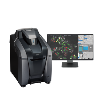
KEYENCE’s All-in-One Fluorescence Microscope BZ-X800 has the versatility to perform fluorescence, brightfield, and phase contrast observations and various imaging methods with a single unit. The BZ-X800 can observe entire well plates, perform quantitative colony counting and area measurement, and solve various problems in experiments, research, tests, and inspections.
This section introduces the advantages of using the BZ-X800 and various application examples.
Observation of an entire well plate using a large motorized stage
In high magnification colony observation using a microscope, it is necessary to repeatedly observe parts of well plates while recording the stage coordinates because the field of view is narrow at high magnification. It is impossible to view the entire well plate and observe fine colonies at high magnification at the same time. In some cases, to acquire an image with a wide field of view, high magnification images are stitched manually into a single image using image processing, which is difficult and time consuming. One issue when observing an entire well plate is how to observe as quickly as possible without being affected by live cells and bacteria that change with time.
The BZ-X800 has an image stitching function that automatically captures high magnification images while controlling the large motorized stage in the X-, Y-, and Z-axis directions and stitches them together, so it is easy to acquire a high resolution image of an entire well plate. Wide field-of-view observation of an entire well plate and high magnification observation of colony locations can be switched seamlessly, which allows for smooth observation without overlooking any colonies that have formed or losing the observation coordinates.
Additionally, image stitching can also be applied to automatically process multiple wells on a multi-well plate at one time.
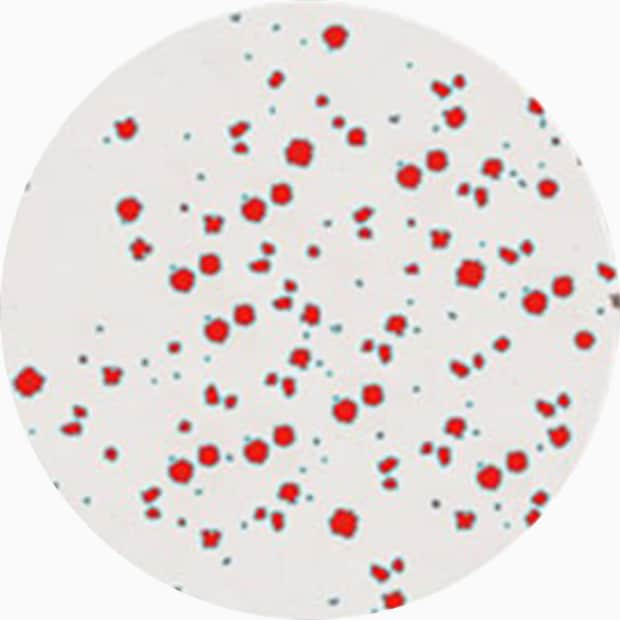
Example of streamlining iPS cell colony counting and area measurement over an entire well plate
Live cells and bacteria change over time, which affects the results of well plate analysis and evaluation, so it is important to obtain quantitative analysis results quickly.
The following images, captured with the BZ-X800, show an example of automatic colony counting and area measurement using Hybrid Cell Count on the image of an entire well plate of iPS cells obtained using image stitching. The number of colonies, average area, standard area deviation, and total area can be measured quantitatively from the wide-field-of-view observation of an entire well plate, which provides analysis results quickly. This can save a huge amount of the time and effort required for manual counting and also eliminates the risk of human error.
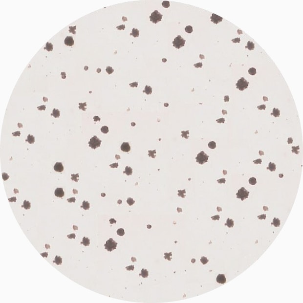
Objective lens: CFI60 CFI Plan Apo λ 10x
Image stitching: 7 images x 9 images
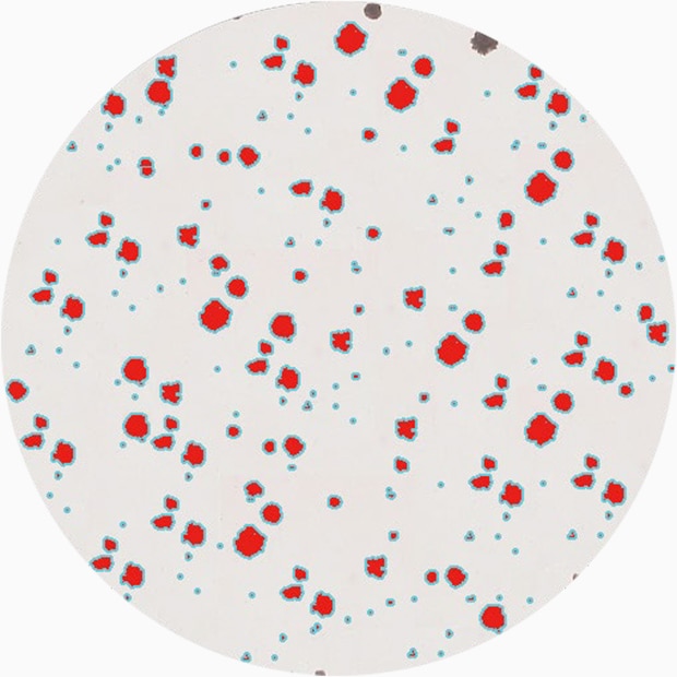
Quantity:251
Average area:8,315 µm2
Standard area deviation:12,652 µm2
Total area:2,086,967 µm2
When a multi-well plate is used, the BZ-X800’s Macro Cell Count automatically processes a batch of multiple images under the conditions extracted for a single image.
Conventional colony analysis over multiple wells requires a huge amount of time and effort, and presents the risk that analysis results and evaluation will vary from operator to operator. With the BZ-X800, quantitative measurement and analysis can be achieved easily and quickly.
Batch measurement under the same conditions

Quantity:181
Average area:5,133 µm2
Standard area deviation:8,730 µm2
Total area:929,027 µm2
Quantity:123
Average area:13,543 µm2
Standard area deviation:17,023 µm2
Total area:1,665,761 µm2
Quantity:171
Average area:12,969 µm2
Standard area deviation:14,976 µm2
Total area:14,976 µm2
Using the All-in-One Fluorescence Microscope BZ-X800
- Image stitching using a large motorized stage automatically captures an entire well plate. Simple operations can be used to observe entire well plates and fine colonies seamlessly at high resolution.
- When a multi-well plate is used, image stitching can be applied to a batch of multiple wells, which makes it easy to acquire an image that encompasses a wide field of view.
- Hybrid Cell Count can perform accurate colony counting and area measurement. Even the area average, standard area deviation, and total area can be measured easily.
- Based on conditions extracted for a single image, Macro Cell Count can be used to process a batch of multiple images. Quantitative analysis can be performed quickly on multiple wells.