Fluorescence Microscopes
Biological Microscopes
What is a Biological Microscope?
A biological microscope is a type of optical microscope that is primarily designed to observe cells, tissues, and other biological specimens. Multiple objective lenses can be attached, which gives these microscopes a magnification that can range anywhere from 10x - 1,000x or more. Since these systems are used mainly to look at very flat samples (e.g. microscope slides, petri dishes, well-plates, etc.), the objective lenses have short working distances and high numerical apertures. Some specialized versions of these microscopes are capable of performing fluorescence imaging and are known more specifically as fluorescence microscopes. Unlike conventional microscopes, biological microscopes are specially designed to create the optimal environment for viewing biological specimens.
Biological Microscope Parts and Functions
The main components of a biological microscope consist of:
-
1Eyepieces – used to observe the image
-
2Objective lens – magnifies the image for easier observation
-
3Stage – used to hold the sample, and often includes clips or other fixturing devices
-
4Condenser – concentrates light onto the specimen
-
5Diaphragm – controls the amount of light reaching the specimen
-
6Light source – used to illuminate the sample
Many modern biological microscopes have made advancements over the traditional components outlined above, including replacing the stage with a mechanical, automated design that can be operated using a mouse. Additionally, some now incorporate digital cameras instead of eyepieces, which allow the image to be projected onto a display screen for multiple users to observe the image at once, and allow for images to be captured, saved, and analyzed further.
How does a Biological Microscope Work?
A biological microscope works by focusing light from the illumination source onto the specimen, via the condenser. The light then travels through the objective lens, and is magnified. Finally, the light passes through the eyepieces, and the image is further magnified and made visible to the operator.
Not all biological microscopes are designed or function the same way, and there are several factors that should be considered when selecting the microscope to use:
-
1Magnification – The magnification range of the microscope should match the size and complexity of the samples being observed. For example, observation of bacteria typically requires high-magnification objective lenses (100x – 150x).
-
2Resolution – The resolution determines the smallest feature that can be resolved. Higher magnification lenses typically offer a better resolution, however advanced biological microscopes can utilize more sensitive light-receiving elements and advanced image processing algorithms to further improve resolution.
-
3Illumination/Observation – As a basic requirement, the microscope should provide uniform illumination and accurate hue representation across the sample. While most biological microscopes are capable of brightfield illumination, more advanced microscopes can also offer phase contrast imaging for easier identification and imaging of cells with low contrast with the background. Additionally, fluorescence imaging can be used to help visualize the biological processes taking place within living organisms.
-
4Operability – The operability of the microscope refers to how easy or difficult it is to adjust the settings to observe the specimen. Basic biological microscopes require settings such as the stage, brightness, and focus to be adjusted manually, typically using knobs or sliders. The most advanced models allow all of the lighting, imaging, and stage settings to be controlled via a mouse or touchscreen, and in many cases the system is capable of automatically adjusting the setup to the optimal settings for the sample.
Biological Microscope Applications
A wide-variety of fields use biological microscopes, including:
-
1Medical research – Biological microscopes are used to study cells and tissues to gain a better understanding of diseases, and their effects on the body. This helps researchers develop new drugs, treatments, and therapies, and understand how the drugs perform under a variety of different variables. Fluorescence imaging can be particularly helpful when performing clinical drug trials, to see how the specimen changes in different environments.
-
2Microbiology – Biological microscopes are used to study bacteria and other microorganisms, to better understand their structure and function, and how they interact with their environment.
-
3Education – Biological microscopes are frequently used in middle and high school to teach students about the structure of cells, and help students gain a better understanding of science in general.
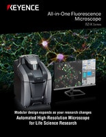
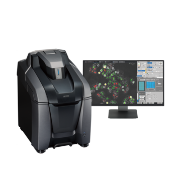
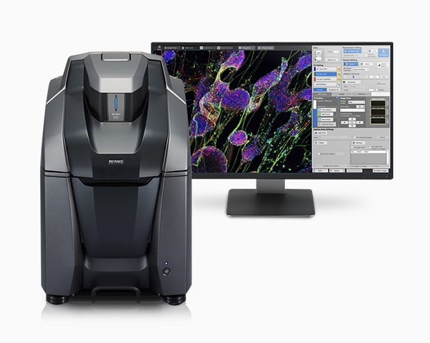
KEYENCE’s BZ-X Fluorescence Microscope
The BZ-X Series is an advanced biological microscope capable of performing brightfield, phase contrast, and fluorescence imaging, and features full electronic operation, so even novice users can capture high-quality images. The system is designed to cover a wide-range of applications, including live cell imaging, cell counting, video capture, and motion analysis.