Digital Microscopes
High Power Microscope
What Is a High Power Microscope?
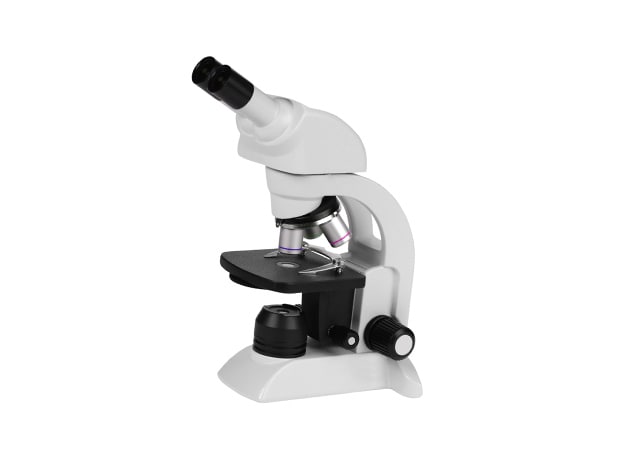
High power microscope is the general term used for microscopes used to observe microscopic targets, such as blood cells and bacteria, that cannot be seen with the naked eye.
These microscopes have a maximum magnification of approximately 1000x. With the standard structure, the light source is positioned below the target to enable observation with transmitted illumination.
The maximum magnification is 1000x because of the limits of optics. Increasing the magnification past this point will not result in better resolution.
(Increasing the magnification past the resolution is referred to as empty magnification.)
High Power Microscope Structure
High power microscopes are also known as compound microscopes.
A compound microscope has a set of two lenses: the objective lens, which magnifies an image of the target, and the eyepiece, which further magnifies the magnified image.
The total magnification of a high power microscope is calculated by multiplying the magnification of the objective lens and the magnification of the eyepiece.
Objective lenses typically have magnifications from 4x to 100x, and eyepieces typically have a magnification of 10x.
High Power Microscope Applications
High power microscopes are also known as biological microscopes. They are primarily used to observe organisms.
They are even used in lessons in schools, with applications including observations of cells and of microorganisms in water.
Limitations of High Power Microscopes
- Short observation distance
- Requires thin samples secured on glass slides
- Normally use transmitted illumination, requiring separate lighting for observation of opaque targets
- Shallow depth of field makes it difficult to observe targets with uneven surfaces
- Capturing images requires an optional camera, but capturing high-resolution images is difficult
- Not equipped with measurement functions because the main purpose is observation
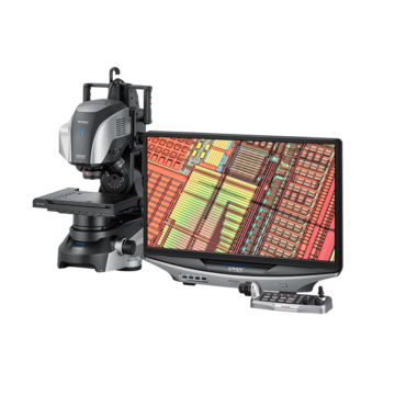
Reasons for Using VHX Series Digital Microscopes
The large depth of field allows the depth composition function to provide observation with the entire image on the screen in focus even at high magnifications
There is no need to cut or polish the target. (Non-destructive observation is possible.)
PCB through-hole, 100x
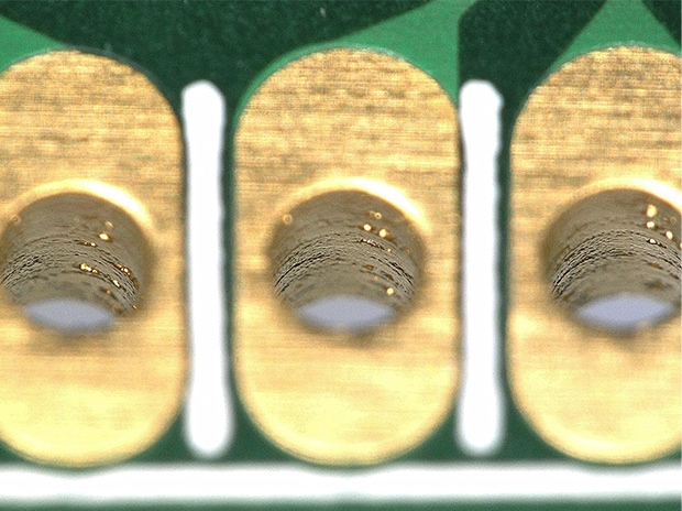
Normal image
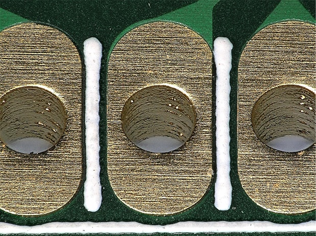
Depth composition image
Being able to select from multiple lighting conditions such as observation with transmitted illumination, ring illumination, coaxial illumination, polarized illumination, and differential interference contrast allows for various applications from biological microscopes to industrial microscopes.
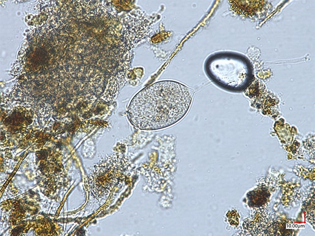
Microorganism in water, 700x,
observation with transmitted illumination
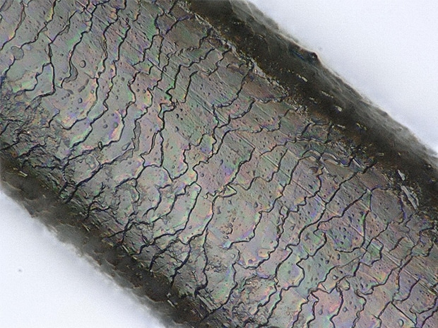
Cuticle, 2000x,
observation with coaxial illumination
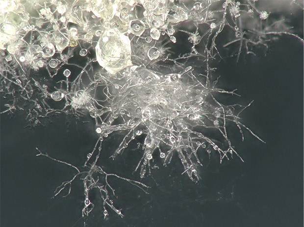
Mold, 200x,
observation with ring illumination
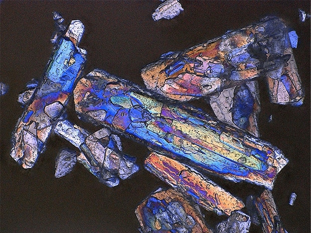
Amino acid crystals, 500x,
observation with transmitted polarized illumination
The standard-equipped camera and monitor allow photographs to be captured with the same quality as monitored images


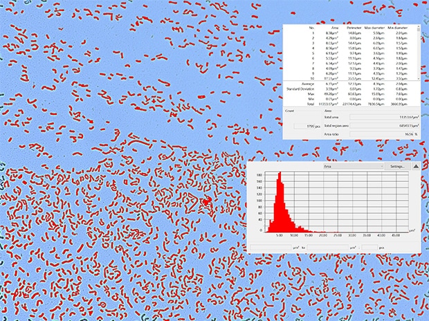
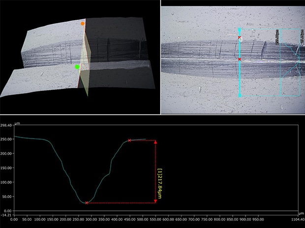
The standard-equipped measuring microscope functions allow for measurement of planes and 3D profiles
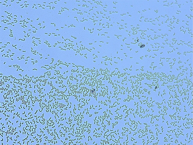
Counting bacteria, 1000x
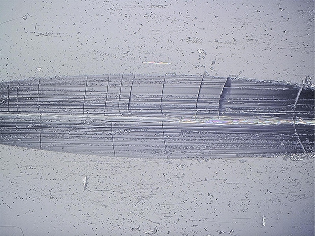
3D profile of the cut-line on an ampule, 300x
![VHX-7000 Series Digital Microscope Catalog [Light version]](/img/asset/AS_129729_L.jpg)