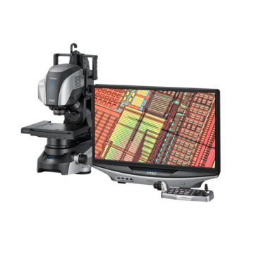
Digital Microscopes
Metallurgical Microscope
What is a Metallurgical Microscope?

A specialized microscope designed for looking at cross-sections of metal targets (metallurgical mounts). Typically inverted, these metallurgical microscopes employ high-resolution objective lenses with very short working distances. While these systems work well for this type of analysis, they are very limited in their range of applications.
Metallurgical Microscopes Parts and Functions
The main components of a metallurgical microscope are the optical system and the illumination system. The optical system typically consists of:
- Eyepiece lens, which is what the observer uses to see the image
- Relay system, which reflects and enlarges the image
- Objective lens, which takes in the image from the sample on the stage
The illumination system is designed to prevent glare from light bouncing off the surface of the sample and consists of:
- High-intensity light source
- Condenser lenses
- Aperture diaphragm
- Plane glass reflector
- Colored or polarizing filters
In some cases, metallurgical microscopes have cameras attached to the eyepiece to capture the images for study.
Uses of Metallurgical Microscopes
Whether they are optical or digital, metallurgical microscopes can be used in virtually any industry or field of study that must observe glossy metal surfaces. These include metallurgy, mineralogy, and gemology. Manufacturers also utilize digital or optical metallurgical microscopes to inspect materials and components for signs of defects or wear.
Limitations of Metallurgical Microscopes
- Objects can only be viewed top-down
- Best-suited for observation in the middle to high magnification range
- Lens magnification is changed with a revolver, cannot change while continuously observing the target
- Mounting attachment is required to add a camera
- Various filters are available to provide multiple observation methods
- Extremely shallow depth-of-field, which means that a defective part may be overlooked
- Short working distance requires samples to be processed before being imaged
- Complicated to operate, requiring users to be thoroughly trained
- Eye pieces cause fatigue
Reasons for Switching from a Metallurgical Microscope to the VHX Digital Microscope
1. Full focus - Sharp focus on the entire target
Digital Microscopes achieve a depth-of-field at least 20 times larger than optical microscopes, allowing for accurate observation of a target with a highly uneven surface.
This type of observation is impossible to achieve with metallurgical microscopes and helps to greatly reduce the amount of time needed for focus adjustments.


2. Grain structure - Quickly analyze grain structure and size on metal surfaces
Different mechanical properties can be obtained even from the same metals depending on the shape, size, distribution, etc of the crystal grains. Therefore, mechanical properties can be determined by examining the state of these crystal grains.


3. Automatic area measurement - Automatically count up to 29,999 particles
The VHX Series’ Automatic Area Measurement function has features that make analysis works more efficient. Images and measurement results can be output into a report format within Microsoft Excel with just the click of a button.


4. Flexibility & hand-held mode -View from any angle, including handheld, to never miss another detail
With Variable-angle Observation capability, digital microscopes can achieve observation from 360-degree angles freely, without having to tilt the target. The VHX Series also offers easy, handheld observation. This means for larger targets, the time required for inspection can be reduced dramatically.


5. 2D & 3D measurement - Perform advanced measurement and analysis
Easily perform 2D and 3D measurements. Roughness, contamination, grain size, and other measurements can be performed with one tool. Data can be saved and accessed later for further measurement. Furthermore, free communication software makes it possible for anyone to utilize the measurement functions from their own PC.


6. 2D & 3D stitching - 2D/3D image stitching and roughness measurement
Digital microscopes can produce fully focused, 3D displays at the push of a button.
Three-dimensional data is obtained while the stage is in motion and images are being continuously captured, creating an overall view of the target. Users are also able to measure surface roughness just by drawing a profile line at any location.


7. Optical shadow effect mode - Delivering images that rival an SEM
Using a specialized design featuring a high-resolution HR lens, a 4K CMOS image sensor and illumination technology, KEYENCE has developed a whole new method of microscopy. Minute irregularities on the surface can be detected by analyzing the contrast in an image captured using multi-directional illumination.


8. Advanced lighting - Automatic acquisition of lighting from all directions
By simply pushing the Optimize button, nine different lighting scenarios are displayed.
From there, you can quickly select the image that is ideal for observing your target.


![VHX-7000 Series Digital Microscope Catalog [Light version]](/img/asset/AS_129729_L.jpg)