Capture publication-quality images right from your benchtop

 Built-In Dark Room
- Install the microscope in any location, so that users can improve their efficiency by eliminating the need to carry sensitive samples between multiple rooms.
 Space-saving
- The all-in-one design of the microscope allows users to place the equipment in areas with limited space or to easily relocate the microscope to accommodate layout changes.
 On Screen Interface
- Everyone can view samples together, increasing collaborative decision making and providing greater confidence in results.
Confocal-grade images without
using a laser
Easily capture high-resolution images without the blurring caused by out-of-focus fluorescent signals. Optical sectioning allows the user to obtain images comparable to those captured on a laser confocal microscope, but in a fraction of the time and without the damaging effects of a laser.
 Conventional
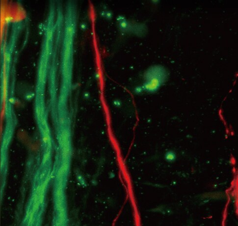 BZ-X SeriesZebrafish
Automated scanning, stitching, and quantification
The BZ-X700 can automatically scan an entire slide or well and quickly stitch together an overall image of a sample. Measurement settings can be stored to and used to quantify over 100 images with the same conditions, providing highly-reliable and repeatable results.
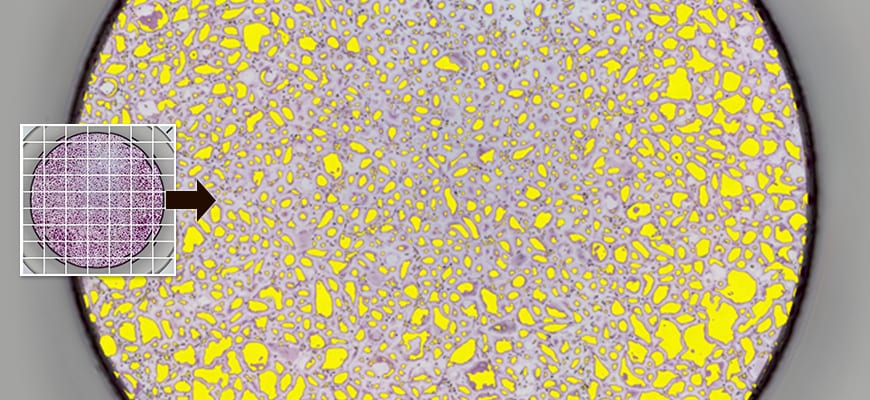 Osteoclasts in 96 well plate
 Brain section
Amazing time-lapse videos without
the hassle
An incubation chamber can be installed inside of the BZ-X700, giving users the ability to perform live-cell experiments and capture time-lapse images. Multiple locations can be analyzed within a single experiment, and the system can produce fully-focused images ,even when at high-magnification, using a Z-stack function. Videos can be recorded at a high-speed of up to 100 fps.
Human Renal Epithelial Cells
Image courtesy of Tottori Institute of Industrial Technology