Fluorescence Microscopes
Quantitative Analysis of Muscle Fibers
-
Tags:
- Clinical Medicine , Neuroscience
Automatic batch measurement
Muscular dystrophy is a heritable muscle disease in which muscle fibers gradually atrophy while destroying and regenerating themselves repeatedly, causing muscular power to weaken.
Pathological findings show that the structure of the bands of muscular fibers will be lost and that affected individuals will have symptoms in muscular fibers such as variability in size, rounded muscular fibers, and increased nuclei.
Objective lens: CFI Plan Apo λ 10x
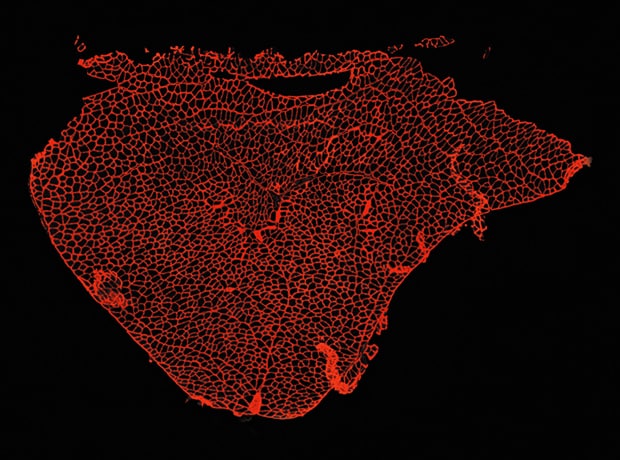
Image stitching: 4 images × 4 images
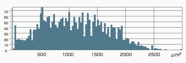
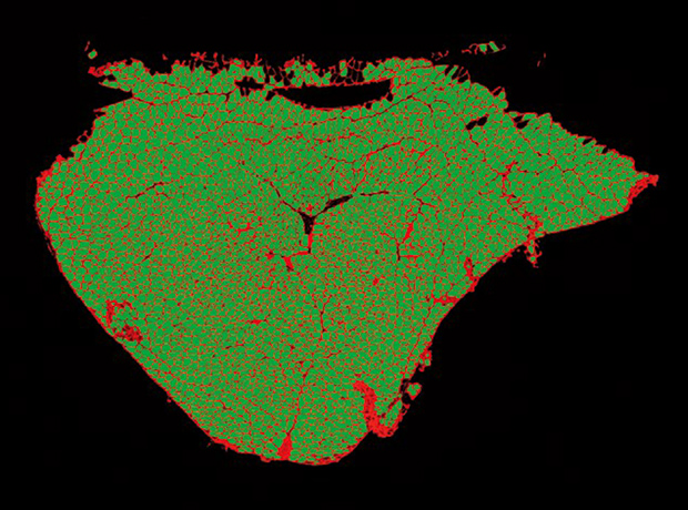
Area of muscle cells
Using the All-in-One Fluorescence Microscope BZ-X800
- When a slice of the target is too large to fall within a single field of view, images are captured while moving the stage and a high-resolution image can be created by stitching these images.
- Even for tilted specimens or specimens that have height differences, it is possible to create an image in which the entire specimen is in focus. This is accomplished by capturing multiple images in the Z direction and stitching together only the parts that are in focus.
- Each closely spaced muscle fiber can be extracted and counted using Hybrid Cell Count.
- On the basis of the conditions extracted with Hybrid Cell Count, the Macro Cell Count can be used to process a batch of multiple images.