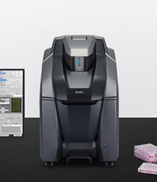
All-in-One Fluorescence Microscope BZ-X800
No darkroom required
Streamline Imaging and Analysis with a Single Platform
01 Enhanced Core Performance
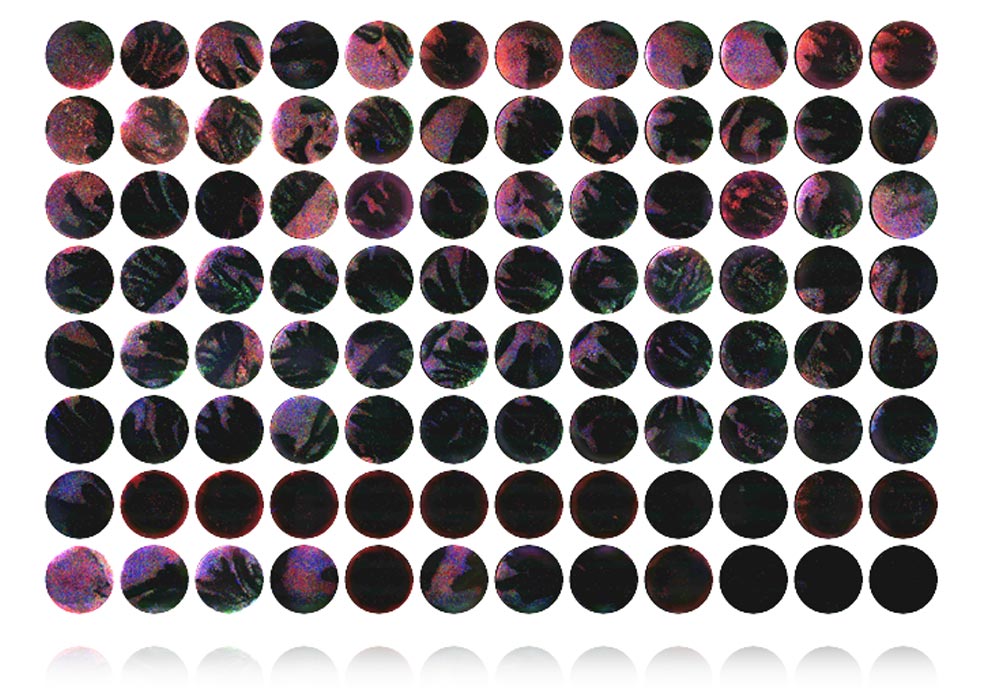
02 Batch Capture and Analysis
03 Scan Slides Instantly
04 Accurate Analysis of 3D Localization
05 Quantify Movement and Changes Over Time
06 Additional Functions
07 Quantification by High-Resolution Stitching Image
08 One-Step Three- Dimensional Quantification
09 Capture Thick Specimens Clearly Using Structured Illumination
10 Fully-Focused High-Resolution, Wide-Area Images
-
 Enhanced Core Performance
-
No darkroom required
Compact size saves benchtop space
-
Full electronic control
Easy operation for all users
-
Publication-quality images
Sensitive optics deliver high-quality results
-
-
NEW
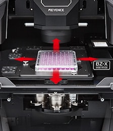
Image Cytometer Module-
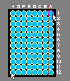
Specify measurement range
-
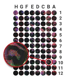
Automatically batch capture desired images
-
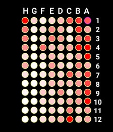
High-content analysis with uniform conditions
-
-
 Scan Slides Instantly
Perform multi-dimensional image capture with high-resolution stitching and Z-stacking.
NEW
Advanced Observation Module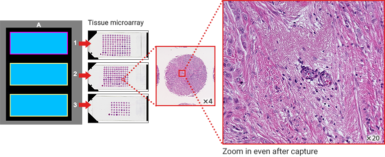
-
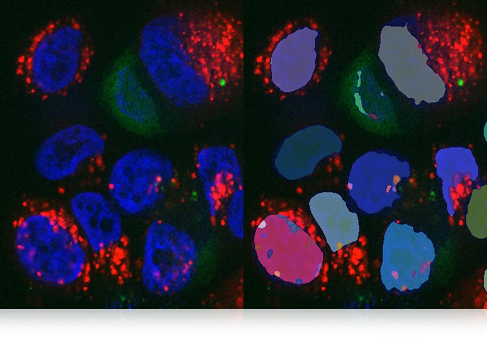
 Accurate Analysis of 3D Localization
Instantly apply quantification conditions to an entire Z-stack. Quantify features such as volume, surface area, and intensity of extracted areas.
NEW 3D Application
-
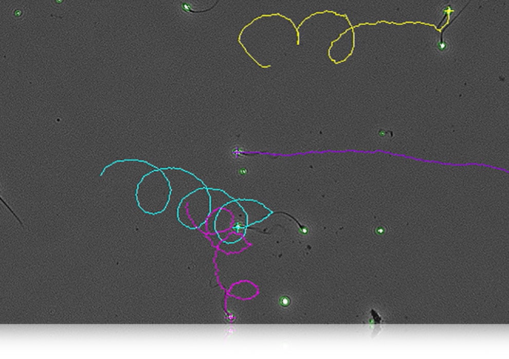
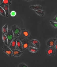
 Quantify Movement and Changes Over Time
Track movement of targets even through morphology changes. Also track brightness, area, and other functions.
NEW Motion Analysis Application
-
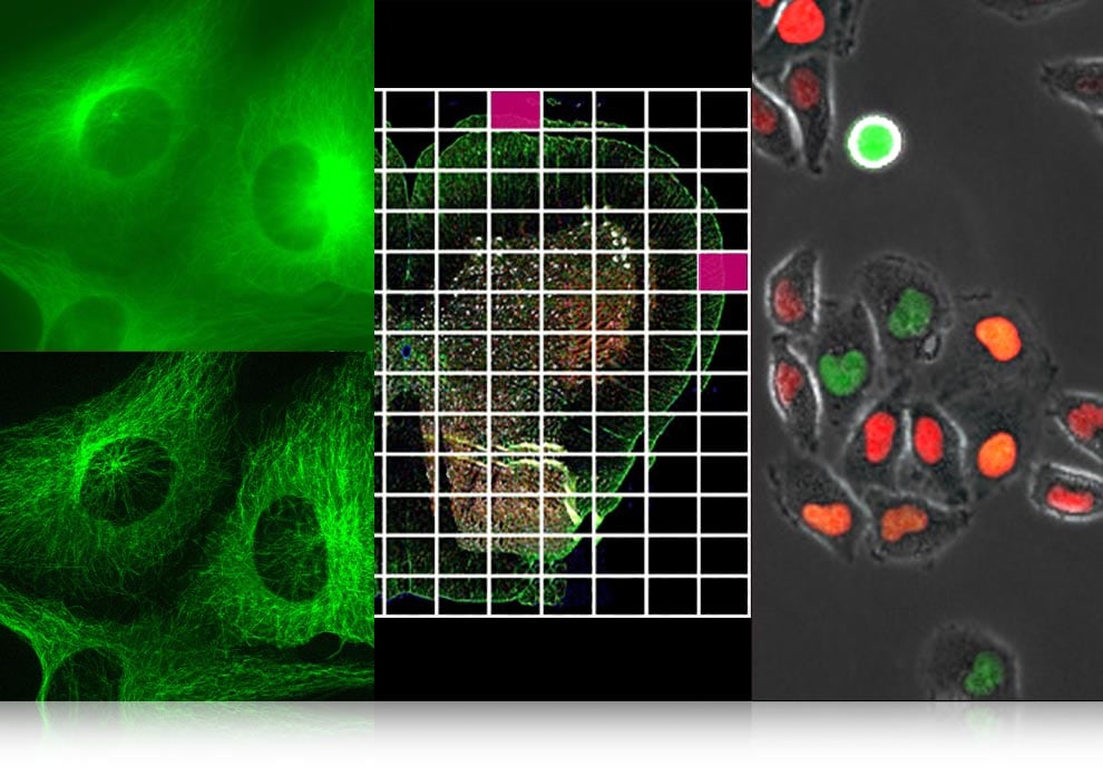
 Additional Functions
-
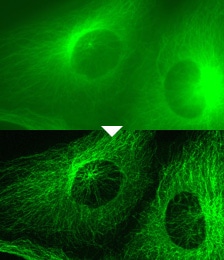
Optical Sectioning
Structured illumination eliminates fluorescence blurring and delivers clear images in just one click
-
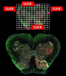
Image Stitching
Capture an entire specimen automatically by registering the coordinates of its outermost positions
-
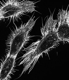
Live Cell Imaging
Accurate analysis of structure and 3D signals
-
-
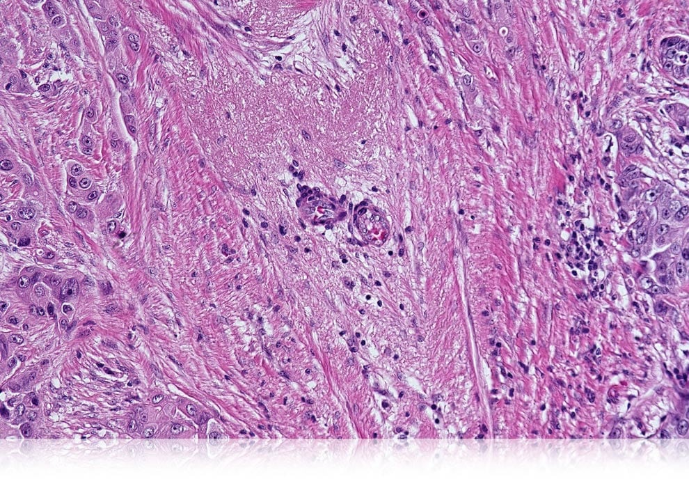
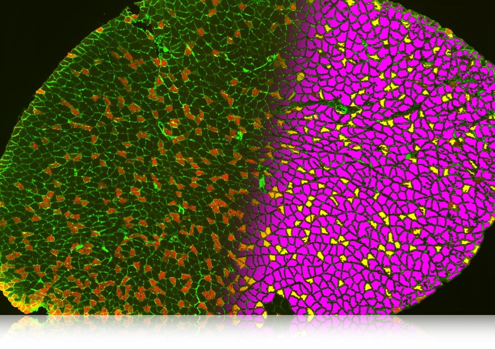
 Quantification by High-Resolution Stitching Image
Slow-twitch skeletal muscle fiber ratio
Muscle fiber 2640 Slow-twitch fiber 540 Slow-twitch fiber ratio 20.5% Courtesy of Lecturer Hideki Yamauchi, Division of Physical
Fitness, Department of Rehabilitation Medicine,
Jikei University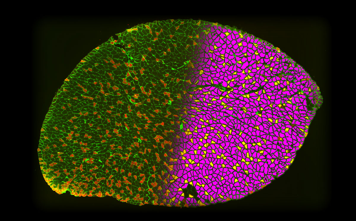
-
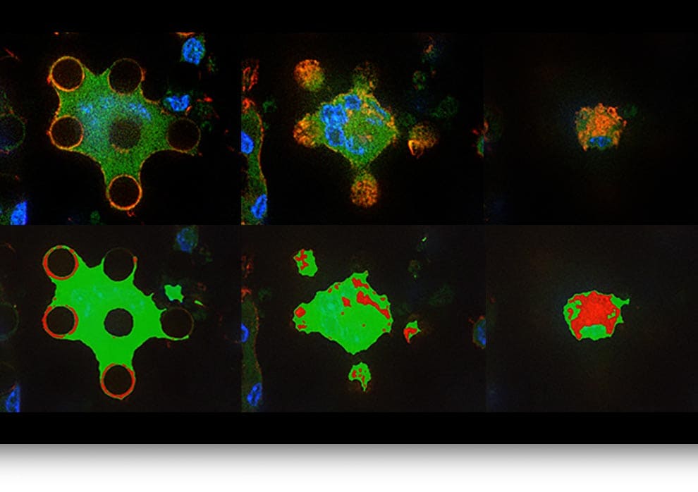
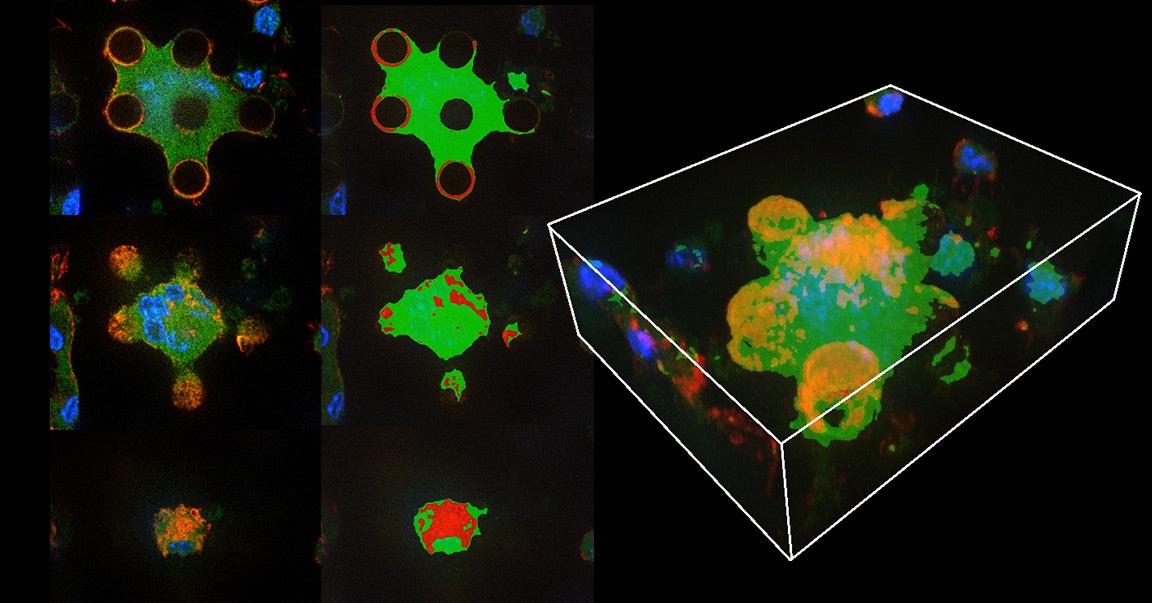
 One-Step
Three-
Dimensional QuantificationMacrophages on nanomaterials
-
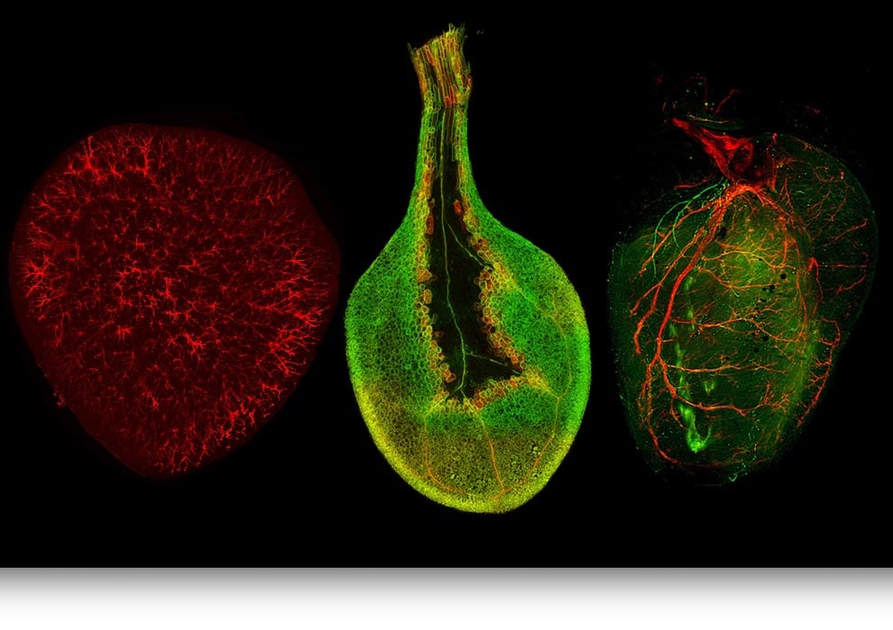
 Capture Thick Specimens Clearly Using Structured Illumination
Courtesy of Dr. Koki Yokoyama, Department of
Cardiovascular Medicine, Osaka University Hospital
Yokoyama et al. PLoS One. 2017 Jul 28;12(7):e0182072.
doi: 10.1371/journal.pone.0182072. eCollection 2017.Cleared specimens
kidney tissue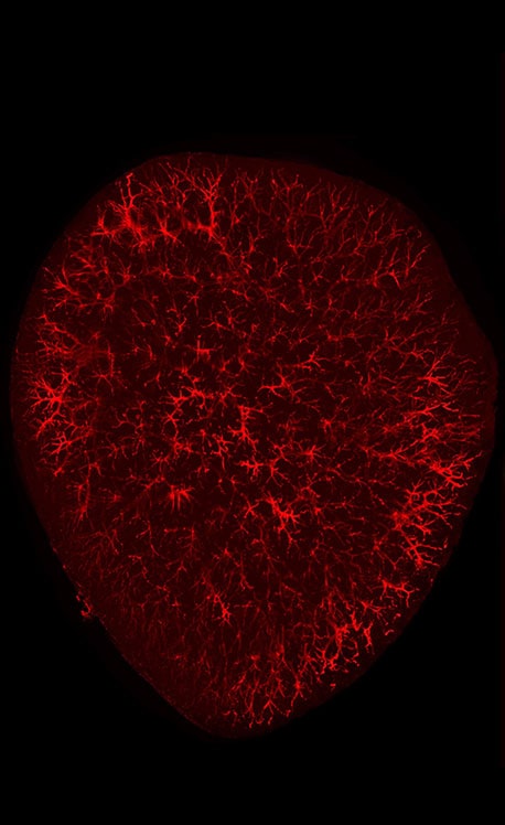
Plant cells
arabidopsis duct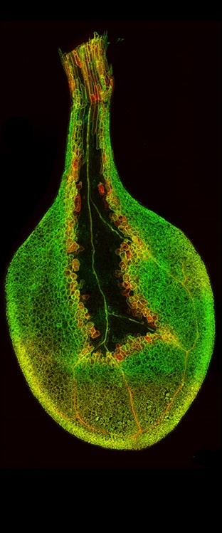
Whole-organ
heart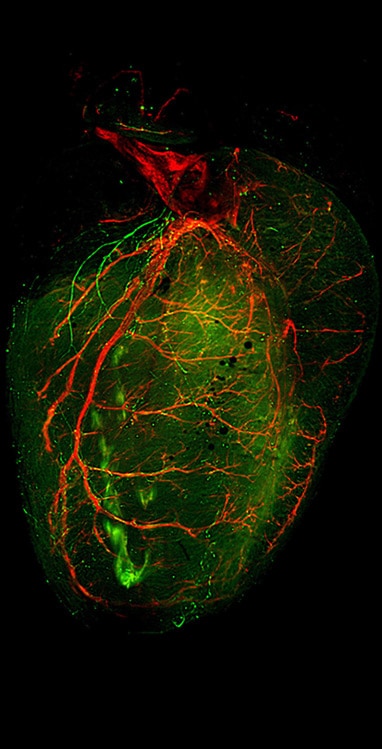
-
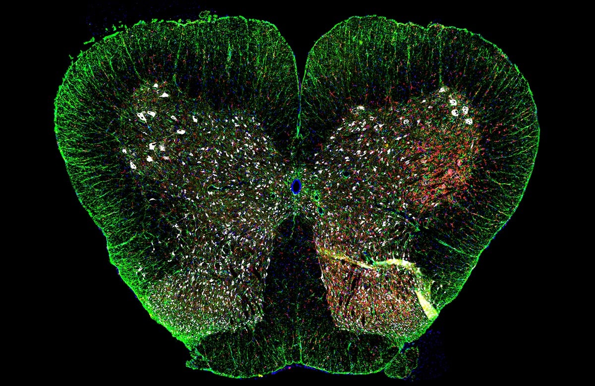
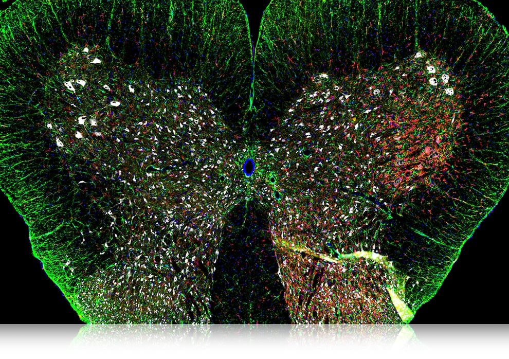
 Fully-Focused High-Resolution, Wide-Area Images
Rat spinal cord
Courtesy of Professor Tasuku Nishihara,
Department of Anesthesia and Perioperative Medicine,
Ehime University Graduate School of Medicine