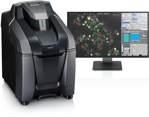
Automated Microscope High-Resolution Imaging and Analysis SystemNEW All-in-One Fluorescence Microscope BZ-X800
High-Definition Images Comparable to Confocal
Structured illumination provides clearer imaging - even on thick tissues - without the damage of a laser.
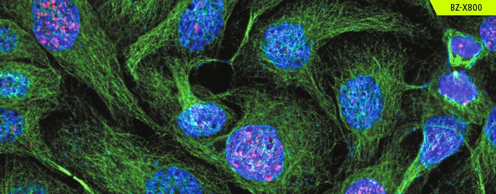
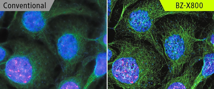
Tubulin and H2AX Courtesy of Momoko Ishikawa, Department of Pediatric Dentistry, Tohoku University Graduate School of Dentistry
High-Resolution, High-Speed Image Stitching
Image an entire slide or well-plate even at high magnification.
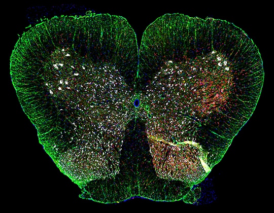
Rat spinal cord Courtesy of Professor Tasuku Nishihara, Department of Anesthesia and Perioperative Medicine, Ehime University Graduate School of Medicine
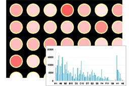
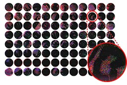
Image Cytometry for Large-Scale Data Analysis
- 01Quickly scan an entire well-plate
- 02High-content analysis with uniform conditions
- 03Automatically batch capture desired images
Capture and analyze in dramatically shorter time frames than with conventional microscopes.
Accurate Analysis of 3D Localization
Instantly convert Z-stacks into 3D images to accurately observe and measure 3D structures.
NEW3D Application
Live-Cell Incubation
Perform time-lapse experiments with ease, including the tracking and quantifying of cell movement.
Courtesy of Postdoctoral Research Scholar Tomomitsu Iida, Leonard Davis School of Gerontology, University of Southern California
When published:Department of Pharmacology, Tohoku University Graduate School of Medicine
View the catalog or application guide for more details.
- You can also contact us at:
