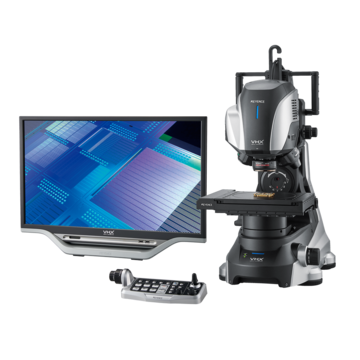
Digital Microscopes
Microscope Observation Modes
Comparison among Observation Mode

A biological microscope uses transmitted light for observation. However, different observation modes have been developed to use specific light characteristics for specific samples, such as transparent specimens and samples that do not pass light.
The table below shows the features of major observation modes. Each observation mode has been developed to properly use characteristics of light such as scattering, diffraction, polarization, interference, and fluorescence.
| Observation modes | Features | Common observation targets |
|---|---|---|
|
|
A general observation mode for biological microscopes, which features a bright field-of-view with transmitted illumination |
Living organisms, cells |
|
|
Uses scattered light, not direct light, to make samples shine against a dark field-of-view |
Microorganisms, cells |
|
|
Uses phase (light variations) to convert a sample to brightness contrast for observation |
Colorless transparent samples, living cells |
|
|
Illuminates a sample with polarized light to convert it to brightness or color contrast for observation |
Crystals such as rocks and minerals, polymers |
|
|
Uses the difference in distances that the transmitted light travels after passing through a sample to generate color or brightness contrast for 3D observation |
Colorless transparent samples, living cells |
|
|
Converts the difference in heights of a sample to brightness contrast for 3D observation |
Cells in plastic containers |
|
|
Stains a sample with a fluorescent compound or fluorescent protein such as GFP to observe the part that expresses fluorescence |
Cells and tissues dyed or labeled with a fluorescent dye, living organisms that exhibit intrinsic fluorescence |
|
|
Uses reflected light to observe samples that do not pass light |
Metals |
|
|
Immerses a sample in index oil and uses transmitted light scattered in the oil for observation |
Detection of asbestos |

Features of Each Observation Mode
Brightfield microscopy
The type of microscopy generally used in biological microscopes. It illuminates the entire sample with direct light and observes it with transmitted or reflected light. This type of microscopy features a bright background and is widely used for inspection of dyed samples, pathologies, and semiconductors.
Darkfield microscopy
This type of microscopy only allows the light scattered or diffracted by the sample to enter the lens so that light can be projected against a dark field of view. It is suitable for observing colorless, transparent samples such as living cells, which are difficult to identify with brightfield microscopy. Darkfield microscopy also enables observation of minute features over the resolution limit of optical microscopes. A special condenser is used for this type of microscopy.
Phase contrast microscopy
This type of microscopy is based on the phase contrast (light variations) caused by light diffraction. Colorless, transparent samples, such as living tissues and cells, are difficult to image using brightfield, but they generate phase contrast due to their differing refractive indexes or thicknesses. Converting this contrast to brightness contrast enables this type of sample to be observed.
This type of microscopy allows cells and other objects that could only be observed against a dark field to be observed against a bright field. In addition, living specimens can be viewed because they need not be dyed. A ring-shaped phase plate with a slit in it needs to be used with this type of observation method.
Polarized light microscopy
Using polarization (biasing of vibration direction of light), this type of microscopy illuminates a sample and makes use of two polarizing plates that can be rotated for observation. If the plates are parallel, the sample appears bright; if the plates are perpendicular, the sample appears dark.
This type of microscopy is useful for observing rock and mineral slices to identify their crystal structures. It is also used to observe fibers, polymers, semiconductors, and bone tissues.
A special objective lens and two types of polarizing plates, polarizer and analyzer, are used for observation.
Differential interference contrast microscopy
As with phase contrast microscopy, differential interference contrast (DIC) microscopy is suitable for observing colorless, transparent samples. It differs in the imaging technique because DIC converts the difference in how light travels to changes in brightness, while phase contrast uses light diffraction.
It is suitable for observing relatively thick samples and also allows for 3D imaging. In addition, because images captured with this type of microscopy complements that captured with phase contrast microscopy, more accurate observation is possible by comparing the results of both.
Objective lenses are marked "DIC" if they are suitable for this type of microscopy.
Modulation contrast microscopy
Also called relief contrast microscopy, modulation contrast microscopy is suitable for observing colorless, transparent samples. Modulation contrast converts the difference in surface heights of a sample to brightness contrast for observation.
As with differential interference contrast, an image can be observed in 3D, although the principles differ. Another difference is that the use of plastic containers is allowed by modulation contrast microscopy, but not by differential interference contrast. Accordingly, this microscopy is suitable for observing sperm and egg cells.
Reflected light microscopy
This type of microscopy uses reflected light for observing samples that do not pass light, such as rocks and minerals. This category can also be further divided into subfields such as brightfield, darkfield, and differential interference.
Dispersion staining microscopy
By immersing a sample in index oil, dispersion staining microscopy performs observation by detecting light dispersions by the different refractive indexes of oil. It is mainly used to identify asbestos and their types.