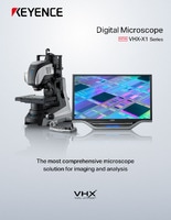
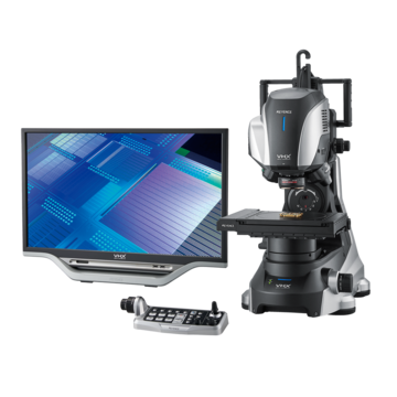
Digital Microscopes
History of Microscopes
Advancement of Optical Microscopes
In the late 17th century, Antoine van Leeuwenhoek of Holland created a simple single-lens microscope. Similar to a modern magnifier in structure, this novel invention differed in that its magnification reached more than 200 times. This new microscope allowed Leeuwenhoek to discover microorganisms and spermatozoa.
Around the same time, Robert Hooke of England created a compound microscope that had two lenses, objective and ocular lenses. Hooke observed cork tissue and called it cells because it looked like the small cells of honeycomb. Thus he coined the biological term cell.
At that time, combining two lenses adversely affected the accuracy, partly due to the aberration of the lenses, resulting in a lower resolution than a simple microscope.
In the 19th century, microscope resolution was improved dramatically through various measures. Aberration correction was implemented by using better lenses or combination of lenses and was supported by German companies such as Zeiss and Leitz who were the main contributors. Ernst Abbe of Germany made theoretical and technical microscope innovations, and it can be said he established the prototype of the modern optical microscope.
Various observation methods were invented in the 20th century. Both the phase contrast microscope introduced in the 1930s and the differential interference contrast microscope invented in the 1950s, contributed to high magnification of transparent samples such as cells. The confocal laser microscope, also invented in the 1950s, marked the beginning of observation with clearer images. Fluorescence microscopes have evolved together with the development of fluorescent dyes around the early 20th century.
Despite these significant improvements in microscopy, 19th century researcher George Airy discovered a limit to resolution based on the nature of light. Soon after, Ernst Abbe introduced the concept of numerical aperture and proved microscopic samples can be resolved only up to 200 nm under visible light, regardless of the performance of the lens, which created a new challenge for magnified observation.

Development of Electron Microscopes
X-rays and the electron were discovered consecutively in the late 19th century, with an electron lens theory introduced at the end of the 1920s. The development of higher resolution microscopes in the early 20th century is a result of using these short wavelength beams as the light source. The transmission electron microscope (TEM) was invented by Ernst Ruska of Germany in the early 1930s, and the first commercial TEM was developed by Siemens in 1939. The development of the scanning electron microscope (SEM) started around the same time as that of the TEM. Next, was the scanning transmission electron microscope (STEM), which was developed by Manfred Ardenne in the late 1930s, followed by the development of the prototype of the modern SEM by Vladimir Zworykin in the early 1940s. Zworykin's SEM, however, had a low resolution, so SEM development continued through projects at Charles Oatley's laboratory at the University of Cambridge in the 1950s, leading to the first commercial SEM produced by Cambridge Instruments in 1965.
Electron microscopes surpassed the limitations of optical microscopes and dramatically improved the resolution so that it is possible to observe objects as tiny as an atom.
In addition to improvements in resolution, many enhancements are still being made to the electron microscope. One is the development of the environmental-scanning electron microscope which maintains the sample chamber at low vacuum for observation of samples containing moisture.