Fluorescence Microscopes
Counting Chromosomes in a Nucleus
-
Tags:
- Pathology , Cancer Research
Capturing clear images without fluorescence blurring
FISH (Fluorescence In Situ Hybridization) is a method of detecting the distribution or amount of specific nucleic acids (DNA or RNA) using fluorochromes. It is mainly used for detecting chromosomal abnormalities.
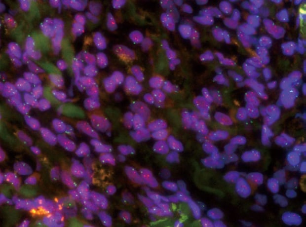
Normal observation
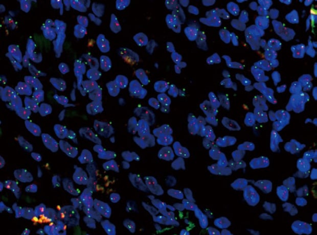
Sectioning + Z-stack
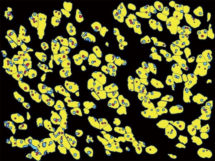
Hybrid Cell Count
Objective lens: CFI Plan Apo λ 100xH
Sectioning + Z-stack
Using the All-in-One Fluorescence Microscope BZ-X800
- The Sectioning function makes it possible to eliminate fluorescence blurring optically and capture clear images.
- A built-in Z-stack function captures multiple images at different focal positions and is able to create a fully focused image by combining only the areas that are at their sharpest focus.
- Hybrid Cell Count can be used to specify nuclei as mask areas to extract and count chromosomes contained in each nucleus.

No. of nuclei containing respective signals
| Signal G\R |
0 | 1 | 2 | 3 | 4 |
|---|---|---|---|---|---|
|
|
40 |
5 |
1 |
1 |
0 |
|
|
23 |
35 |
13 |
1 |
0 |
|
|
10 |
24 |
18 |
2 |
2 |
|
|
0 |
5 |
7 |
2 |
0 |
|
|
1 |
3 |
4 |
2 |
1 |