Fluorescence Microscopes
Live Cell Imaging Microscope
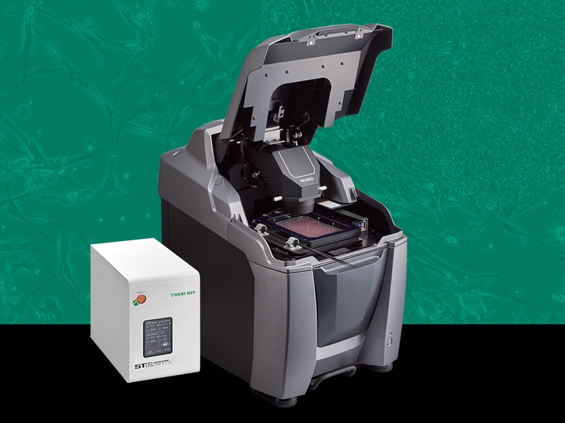
Live Cell Imaging Microscopes are designed to observe the dynamics of living cells in real-time. These systems create and maintain an environment that is conducive to cellular growth, through the careful control of temperature, CO2, and humidity, and are able to capture high-resolution images over time or video in order to analyze cellular structures and functions.
Get detailed information on our products by downloading our catalog.
View Catalog

Control Temperature, CO2, and Humidity for Prolonged Live-Cell Experiments
Our BZ-X All-in-one Fluorescence Microscope integrates an environmental chamber that mounts directly inside of the microscope, allowing users to install the system in any location, including right on their benchtop or in proximity to where specimens are being cultured.
- The small footprint and integrated sample enclosure let you perform time-lapse experiments directly on your benchtop
- Record time-lapse images or real-time video
- Low photobleach mode minimizes cell damage
- Focus tracking ensures clear imaging
- Image multiple locations under different conditions to maximize testing efficiency
- Track changes in brightness and movement over time

Record Time-Lapse Images or Real-Time Video
Record real-time video of cell cultures in fluorescence, brightfield, and phase contrast. The temperature and CO2 regulation chamber can hold a variety of vessels, including well-plates, to create an ideal environment for specimens during prolonged time-lapse imaging.
Minimize Cell Damage
Advanced lighting and camera technology minimizes photobleaching to extend cell life. A high-sensitivity camera is able to capture bright fluorescence images, while the light intensity can be adjusted to accommodate more delicate samples. Additionally, all operations of the microscope, such as focus, position adjustment, or image capture, can be synced with the light source using Low Photobleach mode. This allows for the system to block the excitation light from affecting the sample until it's time to make an adjustment or take a photo.

Coordinate-Specific Condition Settings
Different capture conditions such as focal plane, exposure time, lens magnification, filters, and Z-stack width/step size can be set individually for each registered point. Multiple samples with different conditions can be imaged in the same time-lapse experiment for increased efficiency.
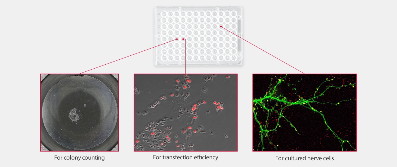
| For colony counting | For transfection efficiency | For cultured nerve cells | |
|---|---|---|---|
|
Lens
|
For colony counting
Phase contrast 10×
|
For transfection efficiency
Phase contrast 20×
|
For cultured nerve cells
Oil immersion 60×
|
|
Observation mode
|
For colony counting
Phase contrast image
|
For transfection efficiency
Phase contrast + fluorescence overlay
|
For cultured nerve cells
Fluorescence 2CH overlay
|
|
Image stitching
|
For colony counting
7×9 images
|
For transfection efficiency
-
|
For cultured nerve cells
-
|
|
Z-stack
|
For colony counting
N/A
|
For transfection efficiency
1.5 μ pitch, 8 images
|
For cultured nerve cells
0.5 μ pitch, 10 images
|
|
Exposure time
|
For colony counting
1/70 s
|
For transfection efficiency
Phase contrast 1/50 s, fluorescence 1/5 s
|
For cultured nerve cells
CH1 1/6 s CH2 1/12 s
|
Focus Tracking Function
The optimal focal plane is automatically selected from Z-stack data. This plane is then set as the center of Z-stack for the next capture to ensure that the sample continues to be in focus. This decreases the number of images captured at each interval, which not only reduces capture time and file size, but also reduces the risk of photobleaching.
Conventional methods
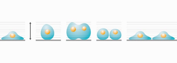
Sets a larger imaging range than necessary to allow for possible movement and morphology changes.
- Larger Z-stack means more images captured
- More exposure to excitation light increases risk of photobleaching
BZ-X
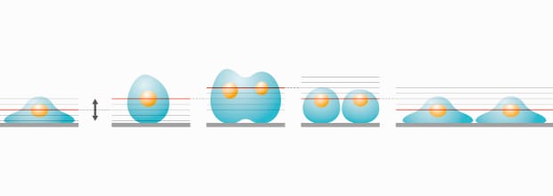
Extracts the optimal focal plane based on previously captured images.
- Less images captured for more efficient review and analysis
- Minimizes sample's exposure to excitation light and reduces risk of photobleaching
Quantify Changes Over Time
This function measures changes in the RGB brightness levels over time. Changes in gene expression can be evaluated quantitatively over the course of the experiment.
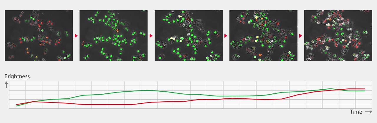
FUCCI cell cycle checkpoints
*Courtesy of Assistant Professor Atsushi Kaida, Oral Radiation Oncology Department, Tokyo Medical and Dental Universitycheckpoints
Track Movement Over Time
Select a target and track it using brightness, hue and appearance information. Automatically record changes in coordinates over time to measure travel range, speed, and movement over time.
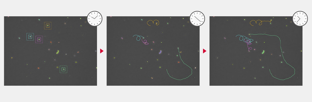
Spermatozoa movementcheckpoints
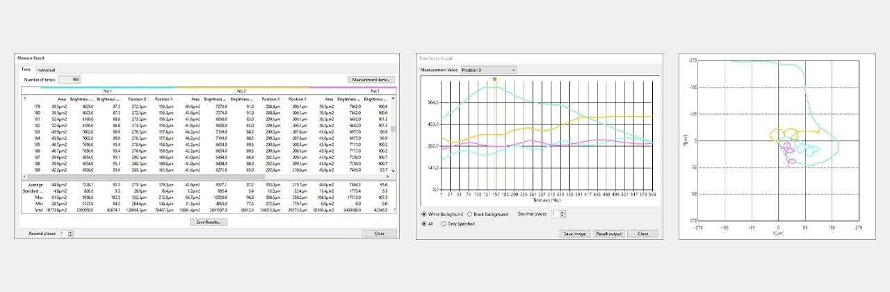
Time-Series Data Output
KEYENCE’s BZ-X Automated Fluorescence Microscope combines many of the capabilities and benefits of both epifluorescence and confocal microscopes into a single, modular platform. At its core, users can quickly and easily capture high-resolution, publication-quality images over wide areas in fluorescence, brightfield, and phase-contrast imaging methods.
This all-in-one fluorescence microscope can be used for routine checks or expanded to accommodate advanced techniques including live-cell incubation and time-lapse imaging, optical sectioning similar to a confocal, low-volume slide scanning, and image cytometry. With an integrated darkroom, users can install this system on any benchtop while taking up minimal laboratory space.
Discover why the BZ-X is the ideal choice for your imaging and high-resolution analysis needs. Download the brochure or request a demo in your lab.