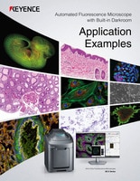
Fluorescence Microscopes
Microscope Slide Scanner
A Microscope Slide Scanner is a high-resolution imaging device used to digitize microscope slides for analysis, storage, and sharing. It captures detailed images of biological samples mounted on glass slides, enabling digital pathology, research, and remote diagnostics.
Get detailed information on our products by downloading our catalog.
View Catalog

Quickly Scan Whole Slides or Tissue Sections in High-Resolution
- Image using fluorescence, brightfield, or phase-contrast
- Focus tracking and Z-stacking ensure images stay in focus throughout the scan
- Combines slide scanner and microscope capabilities for large area, high-resolution imaging
- Quantification software measures and analyzes large data sets
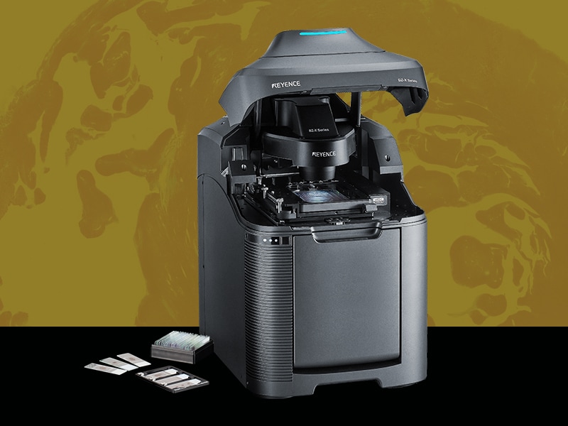
Our BZ-X All-in-one Fluorescence Microscope can rapidly capture a low volume of whole slides in high-resolution while also incorporating all of the functionality of a microscope for more thorough analysis. Contact us to learn more!

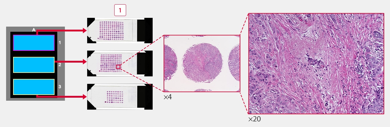
High-Resolution Image Tiling
Simply click the outer boundaries of the area you want to capture and the system will save the XY coordinates, including the focal position, and then quickly scan and capture images throughout that range. See more application images below.
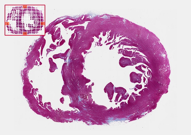
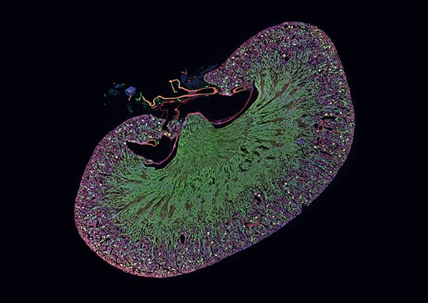
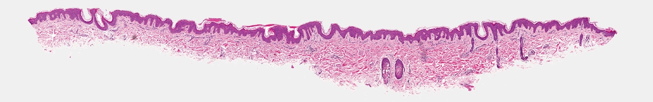
Whole Slide Scans
The BZ-X Wide Image Viewer saves uncompressed images with the highest possible resolution, allowing users to observe fine details of large samples.
-
1Tissue microarray

Eliminate Brightness and Shading Issues on Stitched Images
Our high-resolution optics and advanced software algorithms create a seamless image over your entire sample, without the shading and distortions typically seen with other systems.
Measure and Analyze Large Data Sets
Accurately extract specific areas to measure and automatically apply the same measurement settings across any number of images.
Slow-Twitch Skeletal Muscle Fiber Ratio
Before
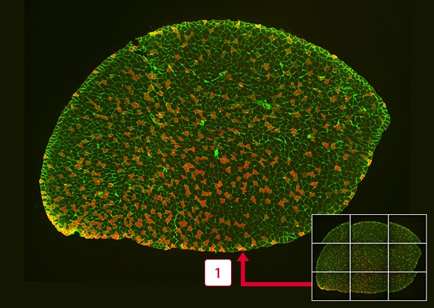
-
1Stitching
After
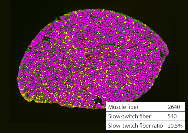
*Courtesy of Lecturer Hideki Yamauchi, Division of Physical Fitness, Department of Rehabilitation Medicine, Jikei University
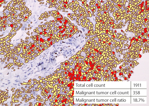
Malignant Tumor Cell (MIB-1) Count
Before
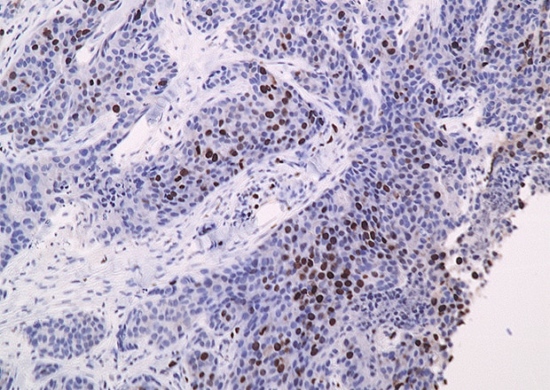
After

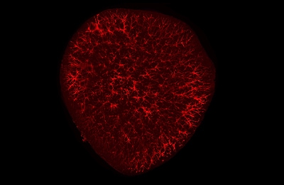
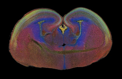
BZ-X Application Examples
*Courtesy of Professor Tasuku Nishihara, Department of Anesthesia and Perioperative Medicine, Ehime University Graduate School of Medicine
*Courtesy of Professor Shigeki Higashiyama, Department of Biochemistry and Molecular Genetics, Ehime University Graduate School of Medicine
*Courtesy of Department of Life Science and Biotechnology, National Institute of Advanced Industrial Science and Technology Masakazu Namihira, Research Group Leader, Biomedical Research Institute
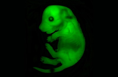
*Courtesy of Lecturer Shingo Nakamura, Division of Biomedical Engineering, National Defense Medical College
KEYENCE’s BZ-X Automated Fluorescence Microscope combines many of the capabilities and benefits of both epifluorescence and confocal microscopes into a single, modular platform. At its core, users can quickly and easily capture high-resolution, publication-quality images over wide areas in fluorescence, brightfield, and phase-contrast imaging methods.
This all-in-one fluorescence microscope can be used for routine checks or expanded to accommodate advanced techniques including live-cell incubation and time-lapse imaging, optical sectioning similar to a confocal, low-volume slide scanning, and image cytometry. With an integrated darkroom, users can install this system on any benchtop while taking up minimal laboratory space.
Discover why the BZ-X is the ideal choice for your imaging and high-resolution analysis needs. Download the brochure or request a demo in your lab.