Fluorescence Microscopes
Human iPS Cell-derived Nerve Cells
Real-time overlay
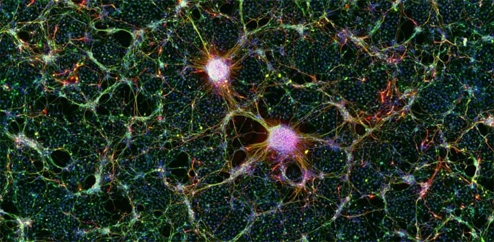
Image courtesy of
Junior Associate Professor Asuka Morizane,
Department of Clinical Application, Center for iPS Cell Research and Application, Kyoto University
The term “iPS cell” is an acronym of Induced Pluripotent Stem cell.
These cells are created by introducing a variety of genes into somatic cells. After a few weeks of cultivation, these cells change to pluripotent stem cells that have the capability to be differentiated into the cells for various tissues and internal organs and the capability to multiply nearly limitlessly.
Using the All-in-One Fluorescence Microscope BZ-X
- Real-time overlay enables multiplex fluorescence images to be overlaid and viewed in real time on the screen.
- Because image capturing conditions can be set separately for each channel, it is easy to optimize the balance of items such as the exposure.
- The image stitching function enables automatic operation that the user executes just by specifying the range to capture. This enables you to easily capture high-resolution and wide-field images.