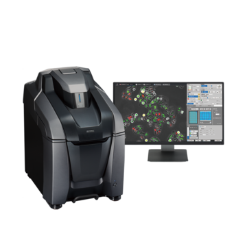
Fluorescence Microscopes
Visualizing Phagocytic Activity (How Microglial Cells Phagocytose Beads)
Capable of stable time-lapse imaging
Source: Courtesy of Postdoctoral Research Scholar Tomomitsu Iida, Leonard Davis School of Gerontology, University of Southern California
(When published: Department of Pharmacology, Tohoku University Graduate School of Medicine)
There are many technical barriers to successful time-lapse imaging, such as focus being lost due to cells moving and reduced cellular activity due to long hours of excitation light emission.
KEYENCE’s All-in-One Fluorescence Microscope can easily perform live imaging thanks to its focus tracking function, pulse excitation, and high-sensitivity camera.
The state of microglial phagocytosis—whether the beads are already taken into a cell or are attached to the surface of a cell—cannot be determined just with phase difference images.
In this case, phagocytic activity is visualized by fluorescent-labeling the beads and using a technique that makes them shine red when they are taken into a cell.
* Specifically, the beads are modified with a pH load (a substance that emits red fluorescence under acidic conditions), which makes the beads red when they are taken into a cell and fuse with acidic lysosomes.
The fluorescent visualization also makes it possible to quantitatively evaluate the cell count and the amount of beads that are taken into cells.