Fluorescence Microscopes
Non-invasive Observation of a 3D Structure Including Rectal Cancer Cells
-
Tags:
- Cancer Research
Easy capturing of clear images without fluorescence blurring
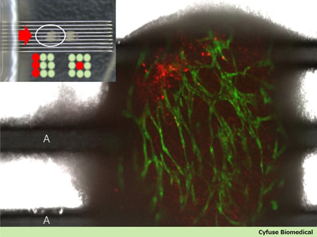
A:Micro needle array
Get detailed information on our products by downloading our catalog.
View Catalog

Points on image capturing
Spheroids were arranged in a 3 × 3 grid in a lamination layer using a Regenova bio 3D printer and were circular-cultured for 6 days. Spheroids fused together, vascular endothelium cells (green) formed a vascular network in intestinal fibroblasts, and metastatic rectal cancer cells (red) grafted to the spheroids also moved. In the yellow parts, cancer cells invaded into blood vessels.

Image capturing conditions
Magnification: 10x λ lens
Sectioning analysis: Captured by using 2D pinholes, at 15 µm intervals, and reclining a 9 × 9 needle array on its side on a tissue culture dish
Filters: GFP, Rhodamine
Fluorescent reagent: Cancer cells already labeled with PKH26 reagent; the image obtained by overlaying 2 types of fluorescence images and a phase contrast image