Fluorescence Microscopes
Cancer Cell Growth
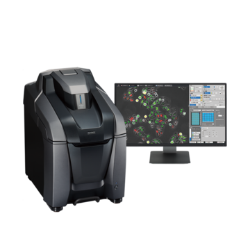
Built-in darkroom, compact design
Video courtesy of
Cell Proliferation Research Group,
Research Institute for Cell Engineering,
National Institute of Advanced Industrial Science and Technology
Get detailed information on our products by downloading our catalog.
View Catalog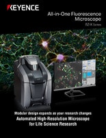

Points on recording
Thanks to a compact design with no need for a darkroom, the microscope can be installed in a cultivation room to perform time-lapse imaging as a routine task. Such an installation eliminates the need to carry targets between rooms and enables the user to obtain high-quality experiment data without concern for contamination or cell damage.

Recording conditions
Magnification: Oil immersion 60x
Glass-bottom dish
1-hour recording in 3-minute intervals