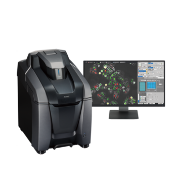
Activity of iPS Cell-derived Nerve Cells
Live cell imaging using time-lapse imaging
Points on recording
By capturing the action potential of nerve cells using calcium imaging, the activity can be easily observed on the basis of visual changes.
Recording conditions
- Magnification: 20x λ lens
- Filter: GFP
- Fluorescent reagent: Fluo-8
- Frame rate: 30 fps
Video courtesy of
Professor Toshiyuki Yamamoto,
Fixed-Term Assistant Professor Keiko Shimojima,
Institute of Medical Genetics,
Tokyo Women’s Medical University
- Here are some examples of using the All-in-One Fluorescence Microscope BZ-X800 in front-line research.
- [Myelodysplastic Syndromes (MDS)] Stitching, Sectioning and the Z-Stack Function as Decisive Arguments for the Acquisition of the BZ Fluorescence Microscope at the University Hospital of Düsseldorf
- [Neuropathology] The perfect solution for everyday patient diagnostics and clinical research at the Institute of Neuropathology in the Charité hospital in Berlin
- [Regenerative Medicine] BZ Series Provides Essential Imaging for Neural Stem Cell and Spinal Observation
- [Gene Therapy] Improving Research for the Development of Gene Therapy Drugs
- [Heart Disease Treatment] Developing Cell Sheets for Myocardial Regenerative Treatments
- [Cancer Treatment] Automated Fluorescence Microscope Transforms Process for Induced Cancer Stem Cell Research
- [Immune System] BZ Series Contributes to Understanding the Pathological Model of Asthma
- [Biomaterials] Promoting Efficiency in Research With Compact, User-friendly Microscopes