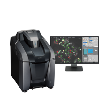
Fluorescence Microscopes
Axonal Elongation of Neural Progenitor Cells (PC12)
-
Tags:
- Neuroscience
High-speed auto focus
Recording conditions
Target cell: PC12 cells in a glass-bottom dish
Lens/Filter: Oil immersion 60x, Semrock GFP filter,
DF + 4% dimmer filter
Get detailed information on our products by downloading our catalog.
View Catalog

Points on recording
Watch the cell in the upper-left part of the image.
The axon from one cell is extending and coming closer to an axon that has extended from another cell.
-
1Even if cells move up and down out of the original focal plane, the BZ is still able to capture a focused image through the use of the Best Focus function.
Conventional auto-focus functions sometimes fail to focus because image capturing is performed according to the relative distance (offset value) from the bottom of the dish. -
2Since these cells are easily damaged by excitation light, reducing the intensity and exposure time is critical. Because the BZ has a highly sensitive CCD, it is still able to capture a clear image with low noise.
-
3Even weak fluorescence signals can be visualized with the Haze Reduction function.
-
4The Multi-point Time-lapse function records, with minimal loss, the “best post-ligand point” that is obtained from a single specimen.