Fluorescence Microscopes
What is Christmas tree staining in sperm inspection?
When conducting forensic investigations of sexual assault, determining if sperm is present on an object or from the victim's bodily fluids is extremely important. Microscopes are commonly used to analyze any evidence that are gathered in order to identify the presence of sperm. There are a variety of methods that can be used to aid in this identification, but in general, it can be very difficult due to the relatively low number of sperm available in a typical sample and the fact that most samples contain a mixture of somatic cells as well.
Christmas tree staining is one method used in microscopy that stains the heads and tails of sperm in a characteristic manner, making it a reliable method for improving identification. Essentially, a Nuclear Fast Red stain solution is used to stain the heads of sperm red and the part of the tip, known as the acrosomal cap, pink. A Picro Indigo Carmine stain solution is used to stain the tails blue and green.
Get detailed information on our products by downloading our catalog.
View Catalog

Improving the efficiency of sperm detection and analysis
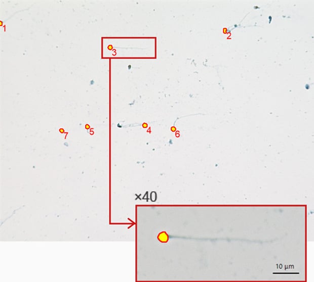
With KEYENCE's All-in-One Fluorescence Microscope BZ-X, it is possible to target the red-stained heads, and then to instantaneously detect and count the number of sperm. Inspection can be automatically done over an entire slide to drastically increase efficiency.
Original image (20x)
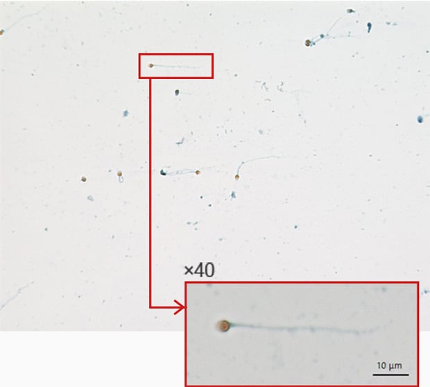
Automatic extraction of sperm head
Automatically scan and capture images over an entire sample
With KEYENCE’s All-in-One Fluorescence Microscope BZ-X, it is possible to capture an image of the entire area of up to three slides with a single operation. Also, the image capture range can be specified by the user, thereby making it possible to save time by excluding unnecessary parts from the captured image.
When using Christmas tree staining, Papanicolaou staining, or other methods where specimens are to be viewed in both brightfield and fluorescence, imaging conditions can be set for each field-of-view to give users flexibility within a single workflow. Some of the conditions that can be adjusted include: illumination method (fluorescence, brightfield, phase-contrast), magnification, brightness, focus, etc.
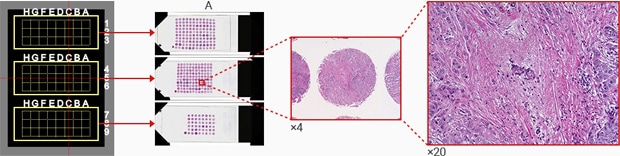
A : Tissue microarray
Quickly identify and measure key features
Several functions available with the BZ-X, such as Hybrid Cell Count and the Image Cytometer module, can be used to automatically detect, count the number of, and measure the area of key regions of a sample based on provided characteristics. These conditions can then be automatically applied across hundreds of images, making it possible to collect data much more quickly and accurately than traditional manual counting methods.
Using the All-in-One Fluorescence Microscope BZ-X
- The motorized microscope stage can be programmed to scan and capture images of multiple slides, dishes, or a well-plate to dramatically increase inspection efficiency.
- Robust measurement software quickly and accurately provides cell count, area, and many other quantitative results.
- The BZ-X requires no darkroom and is fully-motorized, allowing anyone to easily operate the system, even remotely when needed.

