Fluorescence Microscopes
Capturing clear images without fluorescence blurring
Due to the strong effects of fluorescence blurring, axons can be one of the more difficult types of specimens to observe.
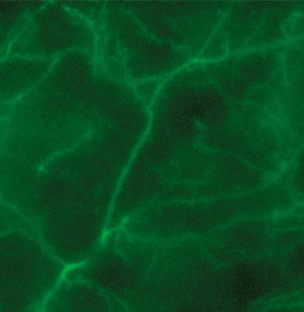
Normal observation
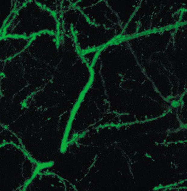
Sectioning observation
Objective lens: CFI60 CFI Plan Apo λ 60x
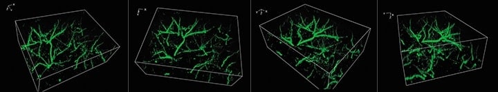
Sectioning + Z-stack
3D image construction
After images are captured at different focal planes, these images can be combined and displayed in 3D with a single click.
Users can then rotate, zoom in or out, and analyze cross-sections of the 3D image to determine the fluorescent signal localization.
Using the All-in-One Fluorescence Microscope BZ-X
- The Sectioning function makes it possible to eliminate fluorescence blurring and capture clear images.
- A built-in Z-stack function captures multiple images at different focal positions and is able to create a fully focused image by combining only the areas that are at their sharpest focus.
- After capturing the focus data at each height, high-resolution 3D images can be created by incorporating the Z-pitch that was used. This helps to give a better understanding not only of the three-dimensional structure of the axon but also of the localization.


