Fluorescence Microscopes
Ultraviolet radiation has beneficial effects on the human body, which include sterilization, promotion of the metabolism and adjustment of biorhythm, and production of Vitamin D to help bone health. In recent years, however, the increase in ultraviolet radiation from the sun due to ozone depletion has been pointed out as a cause of health hazards such as skin cancer and aging caused by sunburn.
This page explains what ultraviolet radiation is, the impact it has on skin, and the mechanism of its effects on skin. Observation and quantification of skin damage caused by UV radiation is also explained.
Get detailed information on our products by downloading our catalog.
View Catalog

What is ultraviolet radiation?
Ultraviolet radiation from the sun that reaches the Earth’s surface (electromagnetic wave) has wavelengths between 100 nm and 400 nm. Johann Wilhelm Ritter discovered ultraviolet radiation in 1801. Its wavelength is shorter than that of visible light—between 400 nm and 780 nm, with violet light having the shortest wavelength (400 nm)—which is where the name ultraviolet comes from.
About the wavelength: In this section, wavelength is explained using values based on photomedicine and photobiology.
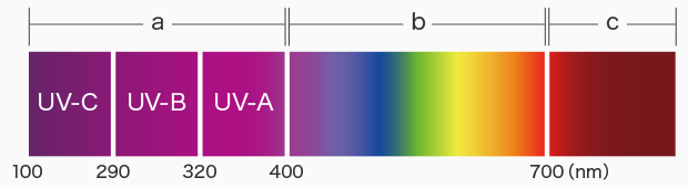
a: Ultraviolet b: Visible c: Infrared
Different bands of ultraviolet rays
Ultraviolet rays are broken down into UVA, UVB, and UVC based on how they affect living things.
UVA (Longest wavelength UV): UV rays with wavelengths of 320 nm to 400 nm.
UVB (Medium wavelength UV): UV rays with wavelengths of 290 nm to 320 nm.
UVC (Shortest wavelength UV): UV rays with wavelengths of 100 nm to 290 nm.
Of the sun’s rays that reach the surface of the Earth, ultraviolet rays account for 5% to 6%, of which approximately 90% is UVA and the rest is mostly UVB. UVC is mostly absorbed by the ozone layer and does not reach the surface of the Earth. Therefore, only UVA and UVB are considered in terms of damage to the epidermis.
Ultraviolet wavelength and photon energy
Ultraviolet is both an electromagnetic wave and light. Light is made of elementary particles called photons, and the energy these particles carry is called photon energy. Photon energy is directly proportional to a photon’s electromagnetic frequency and is inversely proportional to its wavelength. Photon energy E can be expressed by the following equation, where h is the Planck constant (6.626×10⁻³⁴ Js), c is the speed of light, and λ is the photon’s wavelength:
E = h × c / λ
From this equation, you can see that as the wavelength λ get longer, the photon energy E decreases. Conversely, as wavelength λ gets shorter, the photon energy E increases. The force that destroys the molecular bonds in a substance becomes larger as the photon energy increases. The longer the photon’s wavelength, the higher the rate of light reaching the surface of the Earth.
Since UVB has shorter wavelengths, most of it does not reach the surface of the Earth, and thus does it not penetrate the skin to the dermis and damages the epidermis instead. It can cause serious damage in short periods due to its high photon energy.
On the other hand, UVA has longer wavelengths and can mostly reach the surface of the Earth, which also allows it to penetrate the skin as deep as the dermis. Due to its low photon energy, UVA is less likely to cause damage to the skin in short periods, but can damage the dermis if the skin is exposed over long periods of time.

The skin’s protective function against UV radiation
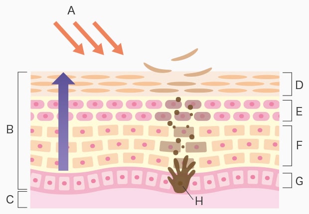
A: Ultraviolet B: Epidermis C: Dermis D: Cornified layer E: Granular layer F: Spinous layer G: Basal layer H: Melanocyte
Skin uses its cell turnover to protect the dermis and hypodermis from UV radiation.
The skin is made up of the epidermis and the dermis. The epidermis is the outermost layer. The epidermis consists of the stratum corneum (cornified layer), the granular layer, the spinous layer, and the basal layer, in order from the surface of the skin downward.
When UV radiation passes through the stratum corneum, the melanocytes in the basal layer generate melanin pigments in order to protect the dermis from UVA and UVB. Melanin is brown or black pigment. The generated melanin pigments are secreted to the keratinocytes in the epidermis and pushed up to the stratum corneum.
Once pushed up to the stratum corneum, the melanin and keratin block UVA and UVB. This process of the skin protects the basal layer and the dermis from UV radiation.
Skin damage caused by UV radiation
As mentioned earlier, the skin has a function that protects the epidermal basal layer and the dermis from UV radiation. However, the skin can still suffer damage from UV radiation when the exposure exceeds its protective function, in terms of the amount of UV radiation or long-term exposure. This section explains the typical symptoms of skin problems caused by UV radiation and how they occur.
Skin problems caused by UVB
As UVB has shorter wavelengths and therefore is absorbed by the ozone layer, the amount of UVB reaching the Earth’s surface is limited, which means that its penetration depth into the skin is shallow. However, the high photon energy means that it can cause suntan and sunburn even over short periods.
Sunburn is redness in the skin that occurs as a reaction to UV radiation. It is generally caused by UVB and is also called an erythema reaction. Sunburn occurs several hours after the skin is suddenly exposed to strong UV radiation. The blood vessels in the reddened area dilate and lead to inflammation. The symptom reaches its peak in 8 to 12 hours. This condition persists for a few days before the skin recovers its normal state.
The dilation of blood vessels promotes the generation of cytokines by keratinocytes that absorbed UVB, which results in an inflammatory reaction. In this process, the adjacent pyrimidine bases in a DNA double helix are joined to form pyrimidine dimers, which are lesions to the cells. Skin cells can repair these lesions with a function called nucleotide excision repair. However, sunburn occurs when the amount of lesions exceeds this function or if repair is not successful. Additionally, the proliferation of such damaged cells can result in malignant skin cancers such as melanoma, squamous cell cancer, and basal cell cancer. Suntan is the darkening of the skin caused by UV radiation, and is also called pigment darkening or tanning. There are two phases in tanning: immediate pigment darkening and delayed tanning.
Immediate pigment darkening
Immediate pigment darkening occurs when the reduced form of melanin in the skin photo oxidizes immediately after the skin is exposed to UV radiation. The pigmentation generally disappears after several hours.
Delayed tanning
Delayed tanning occurs when new melanin pigments are generated over an extended period of time approximately two days after the skin is exposed to UV radiation. Delayed tanning lasts more than several months.
Skin problems caused by UVA
More UVA reaches the Earth’s surface compared to UVB. UVA has longer wavelengths and thus can pass through melanin and keratin in the stratum corneum of the epidermis and penetrate into the dermis.
When UVA reaches the dermis, it affects the elastic fibers, collagen fibers, and mucoperiosteum, which results in deterioration of the suppleness and firmness of the skin, causing wrinkles, sagging, and other aging effects (photoaging) and problems in the skin. It can also oxidize existing melanin and darken the skin.
Quantitative analysis of skin damage caused by UV radiation
Skin damage caused by ultraviolet radiation can be assessed by observing the thickness of stained epidermis using a microscope. Measuring the thickness in any part of the skin can present variations in measurements depending on where you measure, which prevents reliable assessments. Additionally, if a sample does not completely fit inside the field of view, it must be split and therefore be observed multiple times. The increased number of observations required more instances of focus adjustment and lighting setup.
KEYENCE’s BZ-X all-in-one fluorescence microscope allows for observation of the entire image. Click on the four outermost points of the sample image to register the coordinates for quick and continuous imaging. This can stitch images up to a resolution of 50,000 × 50,000 pixels. The BZ-X’s precision shade correction feature eliminates uneven light intensity caused by lens aberrations, which helps remove seams from stitched images. This allows users to observe their samples with natural, seamlessly-stitched images. For localized observation, users can simply click anywhere on the stitched image to instantly move to those coordinates and observe the area in high resolution.
Hybrid Cell Count can be used to accurately extract the target area based on color, brightness, and contour information in order to measure the area. The use of stitched images not only ensures accuracy, but also offers quality data that is visually easy to understand.
The Macro Cell Count feature is useful when there are a number of samples, as measurements can be automatically performed in a batch by applying the measurement conditions from the first image. This reduces measurement time, as well as eliminating variation in measurement conditions, thus increasing data reliability.
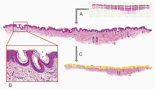
A: Stitching image capture B: Localized high-resolution observation C: Extraction and quantification of damaged site (Area: 72865 μm2)
Example: Observation of mouse skin damage caused by UV radiation
Using the All-in-One Fluorescence Microscope BZ-X
- Image stitching allows for quick acquisition of image data over wide areas. This allows for observation with natural, seamless, high-resolution stitched images.
- The BZ-X can extract the entire epidermal layer for area measurement, thereby acquiring quantitative data quickly and without variations.
- Even when there are many samples that need to be examined, the BZ-X can analyze all samples with the same conditions, achieving accurate comparative assessment while eliminating arbitrariness.

