Fluorescence Microscopes
High-resolution images
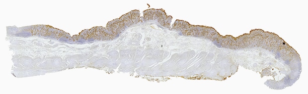
Immunostaining, which is also called an immune antibody method or immunohistochemistry, is often used for histopathological diagnosis. It binds a specific protein (antigen) to tissue with a substance that specifically reacts to it (antibody) in order to visualize tumors or other target parts.
Hybrid Cell Count
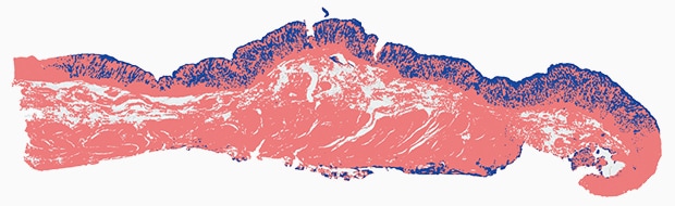
Area (total):65,426,415 µm2
Area (tumor):10,085,121 µm2
Area percentage:15.4%
Objective lens: CFI Plan Apo λ 10x
Image stitching: 20 images × 12 images
Using the All-in-One Fluorescence Microscope BZ-X
- When a slice of the target is too large to fall within a single field of view, images are captured while moving the stage and a high-resolution image can be created by stitching these images.
- Even for tilted specimens or specimens that have height differences, it is possible to create an image in which the entire specimen is in focus. This is accomplished by capturing multiple images in the Z direction and stitching together only the parts that are in focus.
- Hybrid Cell Count can extract only tumoral areas from the entire section and calculate the ratio automatically.


