Fluorescence Microscopes
Fluorescence Microscopy Explained: A Beginner’s Guide
Fluorescence microscopy is an essential tool in modern biological and medical research. By using fluorescent dyes or proteins that emit light when excited, this imaging technique enables scientists to visualize the inner workings of cells with remarkable specificity. Whether you’re new to microscopy or just looking to understand fluorescence better, this guide will walk you through the basics.
What is Fluorescence?
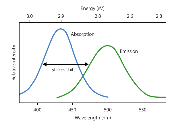
Fluorescence is a phenomenon where certain molecules absorb light at one wavelength (excitation) and emit it at a longer wavelength (emission). This shift is known as the Stokes shift* and is key to detecting specific structures within biological samples.
* The Stokes shift is named after Sir George Gabriel Stokes, a 19th-century British physicist and mathematician. He first described this phenomenon in 1852 when studying the fluorescence of quinine (a compound found in tonic water). Stokes observed that the emitted light had a longer wavelength (lower energy) than the light used to excite the sample — a key insight that laid the foundation for modern fluorescence spectroscopy.
How Fluorescence Microscopy Works
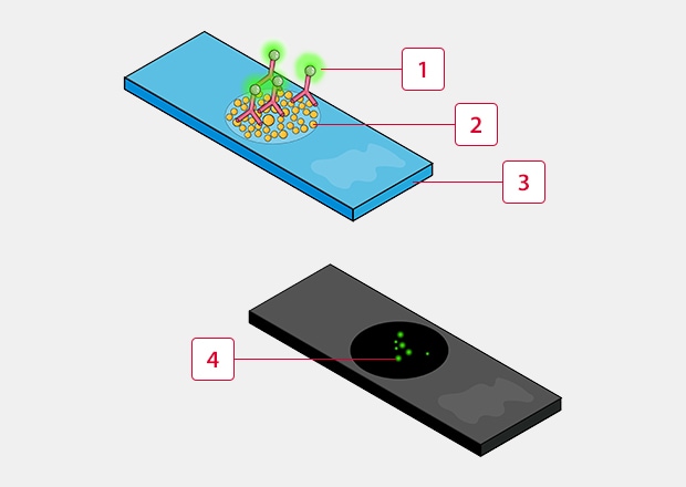
Fluorescent molecules (fluorophores*) are used to tag components of interest within a sample. When the sample is illuminated with light of the appropriate excitation wavelength, the fluorophores emit light that is then collected by the microscope to form an image.
-
1Fluorescent tagged antibody
-
2Taget molecule
-
3Slide
-
4Fluorescence
* While fluorophores are commonly used for tagging samples, there are also several other methods of generating fluorescence, including the use of fluorescent proteins, quantum dots, nanoparticles, etc.
Fluorescence is used to make specific structures in a sample stand out very brightly against a dark background. Here's how it works:
- A fluorescent dye, protein, or molecule is attached to the part of the cell or tissue you want to study.
- When the sample is illuminated with a specific light source (often lasers or LEDs at the excitation wavelength), the fluorophores absorb that energy.
- Almost immediately (within nanoseconds), they emit light at a different, longer wavelength and that emitted light is what the microscope captures.
- Because only the labeled parts fluoresce, you get very high contrast and can see fine details that would otherwise be invisible in regular light microscopy.
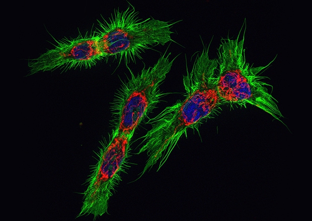
Fluorescently labeled esophageal cancer cells
This method is widely used for studying cells, tissues, bacteria, and even individual molecules, and it's essential in fields like biomedical research, microbiology, and materials science.
The BZ-X All-in-one Fluorescence Microscope captures high-resolution images in fluorescence, brightfield, and phase-contrast across a broad wavelength spectrum – from UV to near infrared.
Get detailed information on our products by downloading our catalog.
View Catalog

Core Components of a Fluorescence Microscope
- Light Source: Typically a high-intensity LED or laser but may also include high-pressure mercury lamps or xenon arc lamps. While lasers can be tuned to a specific wavelength, other light sources are generally broad spectrum and use filters to narrow down their wavelength band. This is needed in order to excite the fluorophores.
- Excitation Filter: Selects the specific wavelength range of light from the light source that is needed to excite the fluorophore and blocks all other wavelengths so that only the desired “excitation light” reaches the sample.
- Dichroic Mirror: A special mirror that reflects the excitation light onto the sample but allows the longer-wavelength emitted fluorescence to pass through toward the detector.
- Objective Lens: Focuses the excitation light onto the sample and also collects the emitted fluorescence. High-quality objectives with high numerical aperture (NA) are often used for better light collection. These lenses are also the primary component used to magnify an image.
- Emission Filter: After the emitted fluorescent light passes through the dichroic mirror, the emission filter ensures that only the specific fluorescence signal (emission wavelength) is captured, blocking any remaining emitted light.
- Camera/Detector: Captures the fluorescent signal and forms the image. These can be simple eyepieces (for visual observation) or cameras (for digital imaging), like CCD or sCMOS cameras for high sensitivity and resolution.
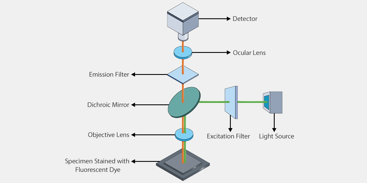
Example of fluorescence microscope internal configuration
The BZ-X All-in-one Fluorescence Microscope incorporates a cooled, monochrome CCD camera with objective lenses ranging from 2x – 100x magnification, including oil-immersion and phase-contrast lenses, and up to four fluorescence channels.

Types of Fluorescence Microscopy
There are several types of fluorescence microscopes, each designed for different levels of resolution, sample thickness, and imaging needs. This is not an exhaustive list, but it does cover the most commonly used types of technologies.
Widefield Fluorescence
Also known as epifluorescence microscopes, these are the most common type of fluorescence microscope and can range from very basic and low-cost systems for routine, quick checks to more advanced, automated systems for research. An entire sample area is illuminated at once and all emitted fluorescence is collected by the detector. They work best for thin samples but can get blurry for thicker specimens since out-of-focus light is also captured.
Confocal Microscopy
These microscopes use lasers and pinholes to focus on very thin planes of a sample by blocking out-of-focus fluorescence light. By removing out-of-focus light, thicker specimens are able to be imaged very clearly and can even be reconstructed into 3D images through optical sectioning.
Two-Photon (Multiphoton) Microscope
Uses two (or more) low-energy or long wavelength photons to excite the fluorophore simultaneously. Only the exact focal point is excited, which reduces photodamage and allows for deep imaging into tissues (even up to 1 mm deep). This method is common for in vivo imaging and neuroscience research.
Total Internal Reflection Fluorescence (TIRF) Microscope
Excites fluorophores in a very thin region right at the surface of the coverslip (about 100 – 200 nm deep). This technique is ideal for studying processes at the cell membrane, like vesicle trafficking or receptor dynamics.
Light Sheet Fluorescence Microscope (LSFM)
This type of microscope is able to illuminate an entire area of a sample with a thin sheet of light. This reduces phototoxicity and speeds up imaging for large, live samples like embryos or organs.
Super-Resolution Fluorescence Microscopes
These systems utilize advanced techniques to break the diffraction limit of light, allowing imaging at resolutions below 200 nm. Some of these techniques include:
- STED – Stimulated Emission Depletion
- PALM – Photoactivated Localization Microscopy
- STORM – Stochastic Optical Reconstruction Microscopy
The BZ-X All-in-one Fluorescence Microscope is a modular system that can function as a widefield microscope, structured illumination microscope, live cell imaging system, slide scanner, and high-throughput image cytometer.

Fluorescence Microscopy: Illuminating the Path to Discovery
Fluorescence microscopy has revolutionized the way scientists observe and understand biological systems. By using fluorescent dyes and proteins, this technique allows researchers to selectively illuminate and study the intricate structures and dynamic processes within cells and tissues. From basic widefield setups to advanced super-resolution and light sheet technologies, fluorescence microscopy offers a wide range of tools tailored to different research needs. Whether you're exploring fundamental cell biology, tracking disease markers, or imaging live organisms in 3D, fluorescence microscopy provides the clarity, specificity, and versatility needed to drive modern discovery. We’ll dive deeper into fluorescence microscopy, its uses, and applications in future articles!