Fluorescence Microscopes
The Indispensable All-in-One Fluorescence Microscope for High-Quality Antibody Development
Explore case studies from Bioss Antibodies that showcase the product's technical advantages and real-world results.
Antibodies – Essential Core Reagents for Biological Experiments
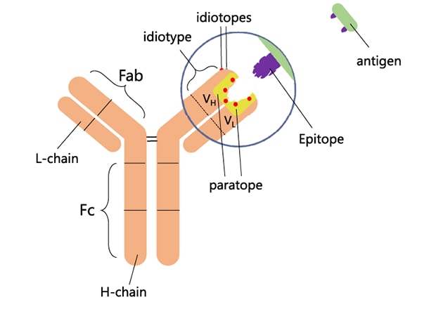
Figure 1-1: Antibody Structure
Antibodies (Abs) are critical tools in modern biomedical research. An antibody is a Y-shaped protein secreted by plasma cells (effector B cells) in response to an antigen, helping the immune system recognize and neutralize foreign substances such as bacteria and viruses. Each antibody recognizes a unique structural feature of a specific antigen.
With the continued expansion of life sciences research, more and more previously unknown proteins have been identified. In studying these proteins, antibodies have become indispensable reagents due to their binding specificity, ease of labeling with enzymes or fluorescent dyes, and broad experimental applications. In biological techniques including, but not limited to, Western blot, ELISA, IHC, IF, and flow cytometry (FC), the choice of antibody is critical. The suitability, specificity, and performance of the antibody directly determine whether reliable, interpretable results can be obtained, making antibodies an essential “core reagent” in biological experiments.
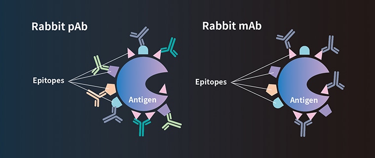
Figure 1-2: Monoclonal (mAb) and Polyclonal (pAb) Antibodies
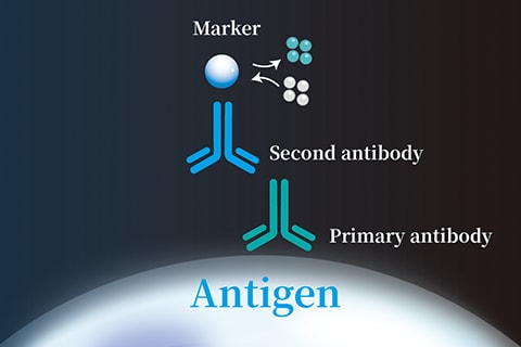
Figure 1-3: Primary and Secondary Antibodies
If you have a background in biology, you may have come across the terms “primary antibody” and “secondary antibody.” Most often, when we talk about antibodies in experiments, we are referring to primary antibodies, proteins that specifically bind to a target antigen. These include both monoclonal antibodies (mAbs), which recognize a single epitope, and polyclonal antibodies (pAbs), which can simultaneously bind multiple epitopes on the same antigen.
By contrast, secondary antibodies are antibodies raised against primary antibodies. Their main role is signal amplification: they bind to primary antibodies and amplify the signal by several orders of magnitude, making the antigen–antibody interaction easier to detect using instruments such as fluorescence microscopes.
Get detailed information on our products by downloading our catalog.
View Catalog

Bioss – A Leading Provider of Immunological Reagents
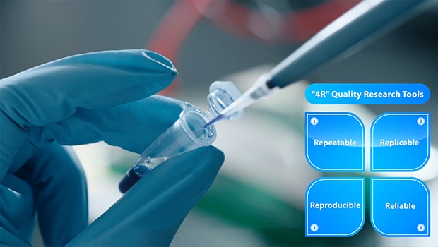
Figure 2-1: Bioss's "4R" Quality Research Tools
Founded in 2001, Bioss Antibodies has been committed to advancing life sciences by providing high-quality immunological reagents and services to researchers worldwide. Backed by a senior team of scientists and advanced antibody discovery, validation, and production platforms, Bioss emphasizes independent R&D and original innovation. Each product is developed to meet international standards and is guided by the company’s “4R” philosophy: Repeatable, Replicable, Reproducible, and Reliable.
With its global brand center based in Boston, Bioss distributes its products to universities, research institutes, and biopharmaceutical companies worldwide, becoming a trusted partner of scientific researchers. While maintaining its commitment to “Made in China, Globally Recognized,” Bioss also strictly follows international quality management systems such as ISO 9001 and ISO 13485.
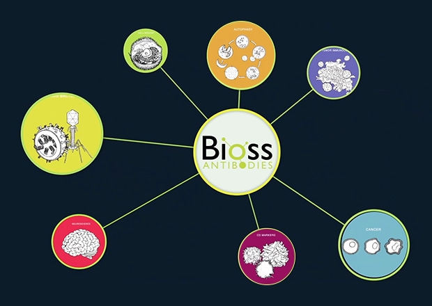
Figure 2-2: Bioss's Application Fields
The company has built a complete R&D pipeline equipped with advanced precision instruments. From target bioinformatics analysis and epitope design to antibody preparation, validation, and labeling, Bioss provides one-stop services that ensure the development of highly specific, sensitive antigen and antibody products. The company also offers industrial-scale platforms for peptide synthesis, purification, and antibody production, capable of generating milligram- to gram-scale outputs. Whether for animal proteins, plant proteins, small molecules, or pathogen-related targets, Bioss has earned broad recognition for its quality and reliability.

BZ Series Microscopes – Supporting Bioss’s R&D Excellence
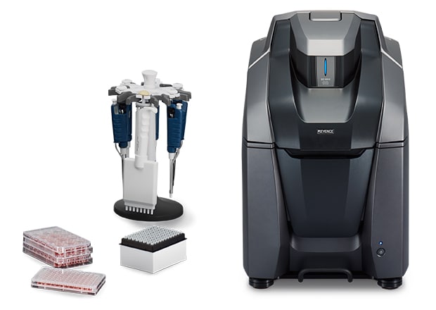
All-in-One Fluorescence Microscope BZ-X
At Bioss’s R&D headquarters in Beijing Yizhuang, Mr. Wang Haoran, manager of the Flow Cytometry Group, shared how the KEYENCE BZ-X800E series All-in-OnE fluorescence microscope has enhanced their antibody research and development.
According to Mr. Wang, the BZ’s highly automated operating system makes observation and image capture simple, consistent, and reproducible, while reducing operator errors. The intuitive interface lowers training costs, and the modular design supports a wide range of experimental needs—including large-image stitching, Z-stack scanning, and high-resolution optical sectioning.

Figure 3-1: Mr. Haoran Wang, Manager of Bioss's Flow Cytometry Group, being interviewed in the lab
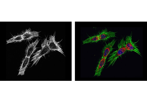
Figure 3-2: BZ's high-sensitivity imaging with a cooled CCD (left) and multi-color fluorescence labeling (right)
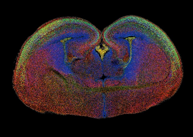
Figure 3-3: BZ's stitched image of a whole brain with multi-color fluorescence labeling
During antibody development, fluorescently labeled antibodies must often be evaluated. For samples with weak labeling and faint signals, longer exposures increase the risk of quenching. The BZ’s cooled CCD sensor enhances the signal-to-noise ratio, while its anti-quenching function prolongs sample life and reduces experimental costs. In comparative experiments, automated recording of all imaging conditions ensures reproducibility and stability across experiments. In addition, the compact design eliminates the need for a dedicated darkroom, saving valuable laboratory space.

BZ Series – Improving Efficiency in Cellular Immunofluorescence
In the field of cellular immunofluorescence, Bioss is committed to achieving both efficiency and quality, which is why the KEYENCE BZ fluorescence microscope has become a central platform in its workflow. Prior to adopting the BZ, the company’s upright fluorescence microscopes required cumbersome preparation steps: cells needed to be plated on slides, and samples had to be sectioned before observation.
The BZ series streamlines this process:
All-in-One Fluorescence Microscope BZ-X
-
1Versatile sample compatibility – Direct observation of samples in common vessels such as well plates, slides, petri dishes, and culture flasks, including both glass-bottom confocal dishes and plastic-bottom multi-well plates.
-
2Fully motorized control – Autofocus, magnification switching, and fluorescence channel selection are achieved with one click. Navigation via the minimap makes it easy, even for beginners,to quickly locate and image cells, reducing training costs.
-
3High-speed optical sectioning – Using structured illumination with LEDs instead of lasers, the BZ achieves high-resolution imaging across a wide spectral range with reduced photobleaching. For thicker specimens, focal-plane information can be accurately captured, producing clear images of animal cells, plant cells, and cultured tissues.
Combined, these features enable faster imaging, lower phototoxicity, and higher reproducibility compared with traditional methods.
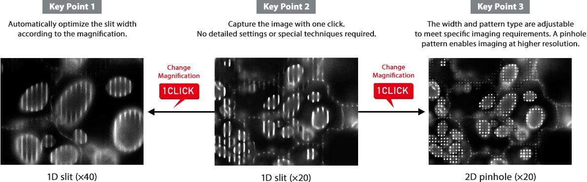
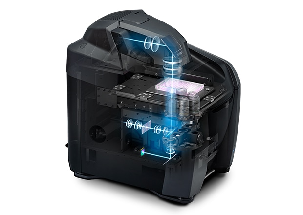
Figure 4-1: Structured Illumination Imaging
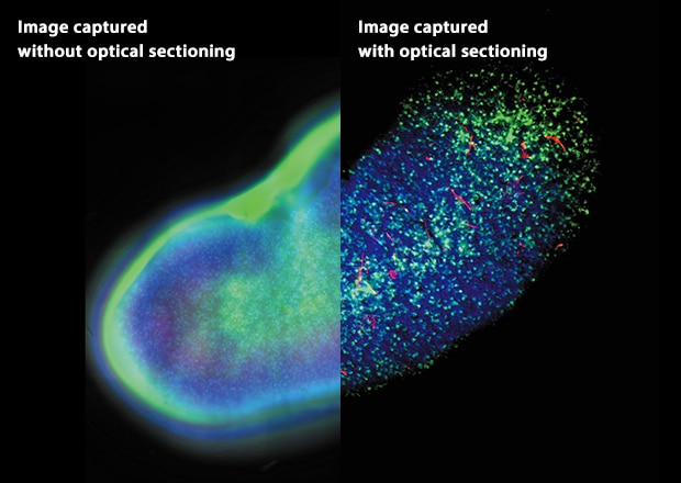
Figure 4-2: Kidney whole-mount (left) and mouse brain neurons (right)
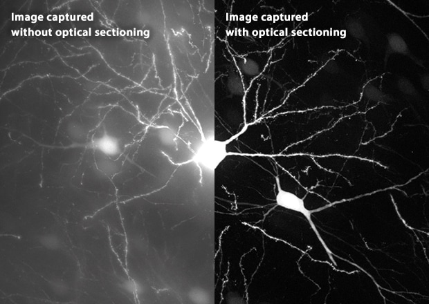

Figure 4-3: A KEYENCE engineer providing imaging guidance
Comprehensive Support
In addition to its advanced hardware, KEYENCE provides extensive pre- and post-sales support. Before purchase, KEYENCE engineers loan the microscope for trial use, helping laboratories evaluate its benefits for their workflow. After purchase, the company offers technical training and rapid-response support for new users or troubleshooting needs, helping enterprises maintain efficiency and maximize their R&D productivity.

