Fluorescence Microscopes
BZ Series Provides Essential Imaging for Neural Stem Cell and Spinal Observation
Dr. Hideyuki Okano
Chairman, Graduate School of Medicine, Keio University
Professor, Department of Physiology, School of Medicine, Keio University
Doctor of Medicine
Dr. Hideyuki Okano was born in 1959. He graduated from the School of Medicine, Keio University in 1983. Beginning as an Assistant to the Department of Physiology, School of Medicine within the same university; he became an Assistant to the Institute for Protein Research, Osaka University; Assistant to the Chemical Laboratory, Institute of Medical Science, The University of Tokyo; Professor of Molecular Neurobiology of Institute of Basic Medical Sciences, University of Tsukuba; and Professor of Neural Function Anatomical Science Research Division of the School of Medicine, Osaka University. He then came to his current post at Keio University in 2001. Since 2007, he has served as the Chairman of the Graduate School of Medicine at Keio University. His main research fields include: molecular neurobiology, developmental biology, and regenerative medicine. Over the years he has been awarded many honors, including the “Medical Award of The Japan Medical Association”; the “Distinguished Scientists Award”; the “Commendation by the Minister of Education, Culture, Sports, Science and Technology (Prizes for Science and Technology)”; the “Stem Cells Lead Reviewer Award”; the “Inoue Prize for Science”; and the “Medal of Honor with Purple Ribbon.” He is also a Board Member of the International Society for Stem Cell Research.
Get detailed information on our products by downloading our catalog.
View Catalog

Researching the generation/regeneration mechanisms of nerve cells for the treatment of spinal cord injuries
Dr. Hideyuki Okano stands at the forefront of the pioneering research field of central nervous system regeneration. With multiple achievements in this field of the world’s highest standards, his research covers much ground. In addition to fundamental research clarifying the mechanisms behind nerve cell generation and regeneration, as an applied study his aim is to establish regenerative treatments for spinal cord injuries by utilizing stem cells. While challenging the common medical perception that the central nervous system of an injured person cannot regenerate, Dr. Okano’s research continues toward the aim of achieving regenerative treatments that are proven to be safe.

01. Ground-breaking research linked with the treatments of nervous system diseases and injuries

One research topic undertaken by Dr. Okano is the establishment of a system effectively derived from various neural cells (neurons) or glial cells (that make up the central nervous system structure) using neural stem cells. The above image is reproduced with permission from the Laboratory Website.
In recent years, the possibility of regenerative medicine has become more of a reality. Talk of ES cells and iPS cells has frequented the news, gathering the interest of the general public. Already, skin regeneration is, to an extent, being utilized practically, and research into the artificial manufacture of blood is advancing.
Within the various regenerative medicine sciences, the field that holds the most expectations while being the most difficult to achieve is the regenerative treatment of the central nervous system: the brain and spinal cord. This stems from the common perception of “once injured, any adult mammal cannot regenerate the central nervous system,” which has been long accepted in the medical world.
The scientist who has destroyed this preconception with his groundbreaking discoveries is Dr. Hideyuki Okano, a Medical Professor at Keio University. Dr. Okano made world history by being the first to discover the existence of neural stem cells in adult brains, providing evidence of the possibility of using these cells in the regeneration of the central nervous system.
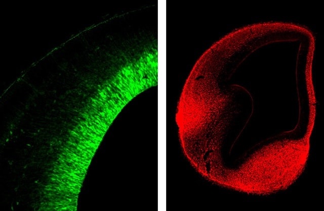
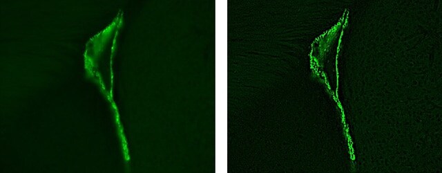
The photograph on the left depicts a neural stem cell, the base of the nervous system. The photograph on the right is a neuron.
(Photographs provided by: Assistant Professor Dr. Takehiko Sunabori, School of Medicine, Keio University)
After researching the differential control mechanisms of neural stem cells using the drosophila fly in the 1990s, Dr. Okano has continued his research through studying the control mechanisms in the neural development of mice and the common marmoset—close in the order of primates to humans. Through his research, Dr. Okano is facing the challenge of solving the mysteries behind generative mechanisms of the central nervous system that use neural stem cells, ES and iPS cells existing in the adult brain.
Neurons and glial cells that make up the nervous system are produced together from neural stem cells. Dr. Okano has not only clarified this process, but by explaining the fundamental mechanisms behind brain cell generation, he is attempting to link these discoveries with the treatment of injuries and disorders of the nervous system. The course of this research is gathering the attention of the entire world.
02. Shedding light on the treatment of spinal injuries once thought to be untreatable
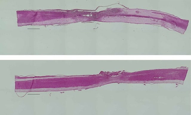
The upper diagram depicts a fibroblastic cell grafted onto a damaged spinal cord. The lower diagram depicts a damaged spinal cord (samples are infused with buffer solution). The overall length is approximately 14 mm. After each sample is stained with H&E, the samples are magnified. The photograph is synthesized using the image stitching function of the All-in-One Fluorescence Microscope BZ Series.
(Photographs provided by: Dr. Osahiko Tsuji, Orthopedic Surgery, School of Medicine, Keio University)
A key target of Dr. Okano’s fundamental research is the treatment of spinal cord injuries. Spinal cord damage can arise from something as simple as an accidental fall or can be caused by disease. Once the spinal cord is damaged, there is no natural regeneration to fix the problem. This can then lead to partial or full loss of motor or sensory functions. According to the Ministry of Health, Labour and Welfare, every year over 5,000 people suffer from spinal damage caused by incidents, such as motor vehicle accidents, within Japan alone. A total of over 100,000 individuals suffer from paralysis.
Dr. Okano’s engagement in the regenerative medicine of the nervous system was triggered by his experience of witnessing one of his former teachers becoming physically disabled due to spinal damage. He then started to challenge an unknown research field from his sense of responsibility as a doctor to find some way to cure the teacher. In the course of this research, after announcing the possibility of regenerative treatment of the central nervous system, he received many letters from patients suffering from spinal cord injuries. This response renewed Dr. Okano’s sense of the sheer enormity and significance of his research.
Currently, research into the medical treatment methods of spinal damage has surpassed the initial steps of fundamental research and is now attempting to head toward the next stage of development.
“Our intention is to face an area never before attempted by anyone by permanently curing intractable spinal patients. It is for this reason that we are attempting to combine the theoretical evidence gained by fundamental research and the translation of this research to the clinical side. Then we plan on putting every effort into ensuring that clinical research using neural stem cells, ES cells, or iPS cells becomes a reality in the near future. To achieve a highly safe method of treatment, we wish to strictly conduct our fundamental research and clarify the mechanisms behind regeneration.” These words from Dr. Okano express his research ambitions.
03. Actively undertaking researcher development and joint research
In addition to advancing fundamental research toward the regenerative treatment of the nervous system, Dr. Okano is also concentrating his efforts toward the fostering of the next generation of researchers. In 2003, the “Center for the Integration of Basic and Clinical Research in Stem-Cell Medicine and Immunology—New developments based on human cells and in vivo experimental medicine” was adopted by the “21st Century COE Program” of the Ministry of Education, Culture, Sports, Science and Technology. Over 5 years, considerable research results of the world’s highest standards were achieved in stem cell biology, regenerative medicine, immunology, and autoimmune disease research.
“This is a field that really stimulates your intellectual curiosity as a researcher, so I’m expecting great things from young scientists.” − Dr. Hideyuki Okano
Although he says “the world of regenerative medicine is a harsh and competitive world for researchers globally,” Dr. Okano also sends a passionate message that “this is new ground that no one has touched, and as a researcher no other work is as enjoyable or as satisfying. I sincerely hope that young scientists with a ‘can do’ attitude can contribute toward this research.”
Furthermore, Dr. Okano is also highly motivated in regards to joint research. He clearly acknowledges, “Other universities and the private sector have achieved a large number of real results in this field. Moving forward, we hope to tackle a better system of research through cooperation.”
Dr. Okano stands at the forefront of the world’s regenerative medicine for the nervous system. His stance holds the key to the possibility of fundamentally altering medical treatments.
04. A laboratory filled with the world’s most prestigious researchers and equipment
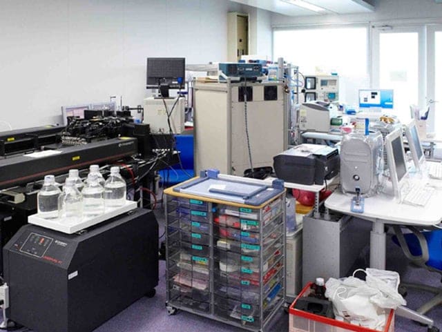
A laboratory scene displaying the line-up of state-of-the-art observation and measurement devices.
Currently, Dr. Okano’s laboratory consists of 5 research groups and 2 independent (professorial) chairs. With a team of researchers, engineers, secretaries, and other staff of 70 strong, this laboratory can be considered world class. In addition to the laboratories that house the common marmosets, mice, or drosophila flies, this research facility stands at the forefront and is equipped with the latest observation and measurement devices, including confocal microscopes and cell sorters.
Of the numerous microscopes being used, the latest to be introduced into the laboratory is the KEYENCE BZ Series fluorescence microscope. Amongst the laboratory researchers, it was said that the assessment of this device was positive during demonstration, even before the device was purchased. One example application for this microscope is the observation of stem cells in relation to spinal damage. This device enables the observation of the entire spinal cord at low magnification.
Additionally, the image stitching function allows for an even larger field of view by merging several magnified images into a single, wide-field image, reducing the amount of time needed to conduct research. By using this function, wide-range images with even brightness can be created in a short period of time by automatically associating multiple images.
Dr. Okano expresses the results of introducing the BZ Series into the laboratory workplace: “Up until now, image synthesis was a troublesome task, taking up much of the researchers’ valuable time. However, by using the BZ Series, an experiment that once took 2 weeks will now only take us 3 days. With this type of increased efficiency, we can repeat the same experiment many times, increasing the throughput. In this manner, we are able to confirm the experiment reproducibility, which is the backbone of accuracy. This is a particularly important component of fundamental research, and a great advantage for any scientist in the pursuit of the truth.”
05. The BZ Series—essential in the observation of the overall image of the spinal cord
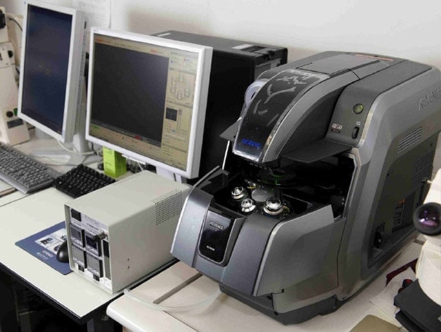
The BZ Series installed within the Laboratory. One advantage lies in this device’s compact size.
Despite only just being introduced into the laboratory, the BZ Series Microscope is already being used by over 20 researchers. This is because the user-friendliness of the various functions links directly with the improvement of research efficiency.
Dr. Okano evaluates the BZ Series: “Simply by conducting research in an unknown field such as this, it becomes of utmost importance to strictly prove whether or not results are showing that nerve cells really are regenerating, or whether the results are a singular phenomenon. For this reason, it is essential that we first use the BZ Series to confirm the entire image of things such as the spinal cord.”
In one of the world’s leading laboratories, the introduction of a large number of state-of-the-art observation devices is not surprising. Amongst all of these devices, according to Dr. Okano, the “BZ Series was essential.” “There are various microscope devices for high-magnification or high-resolution uses made from different manufacturers. However, the conclusion was made that in situations where the entire cell needed to be observed, there was only the BZ Series.”
When deciding on the purpose of this purchase, the general versatility of the device within the laboratory had to be taken into consideration. “The decision to purchase the BZ Series was made after taking into consideration the device’s ability to benefit all research staff and not be limited to a single use.”
In terms of general versatility, the ease with which any person could use the device was also considered. From this standpoint, the BZ Series differs from confocal microscopes in that a high level of skill is not required to use the device, nor does it require a specialist engineer to work with the device. Even a first-time user can quickly become proficient in operating the microscope, adding to the appeal of benefiting the overall research.
In addition to the improvement of research efficiency, Dr. Okano also points out another key feature: image quality. “A clean and clear magnified image can make the difference between a thesis passing or failing its review,” he says. From this perspective, the BZ Series leaves nothing to be desired.
06. Functions unique to the BZ Series contribute to the improvement of research efficiency
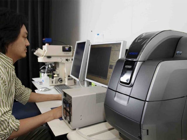
Dr. Takehiko Sunabori,
Assistant Professor, School of Medicine, Keio University
We asked the opinion of a researcher actually using the BZ Series in the laboratory. Dr. Takehiko Sunabori (Assistant Professor, School of Medicine, Keio University), is currently studying the differentiation mechanisms of neural stem cells of the cerebral cortex. He takes a sample by slicing a cross section of the cerebral cortex of a common marmoset. He then uses the BZ Series to capture over time the process of neuron production from neural stem cells.
Dr. Sunabori uses the BZ Series together with a confocal microscope when observing the samples, and he states that “to observe the entire sample at low magnification, or to correct the focus of a blurred image, the BZ Series is extremely beneficial.” He goes on to say that he occasionally uses the image stitching function that corrects for any connecting links or brightness differences when stitching magnified images.
“Up until now, we have manually stitched together individual images. This takes time and there was always uneven contrast, leading to a problem with image quality. This task is now automatically conducted by the BZ Series, thereby resolving all our previous issues. Whereas previously it could take several hours to correct and combine a series of six photographs of the cerebral cortex, now it only takes a few minutes. I am deeply satisfied with the ability to acquire high-quality images.”
According to Dr. Sunabori, in recent years, we are in “an age where good quality is taken for granted” when referring to photographs published within a thesis. Dr. Sunabori says that he himself always pays careful attention to image quality when taking photos.
07. The BZ Series starts up quickly after power-up, enabling immediate observation
The function that Dr. Sunabori truly appreciates when observing the entire image of a sample is the quick full focus function. The focal position of the objective lens is moved electrically along the Z-axis, and images are captured at different focal points throughout the sample, producing a fully focused image. In situations where the depth of field differs, such as the axons of the central nervous system, using this function eliminates the cumbersome task of adjusting the focus.
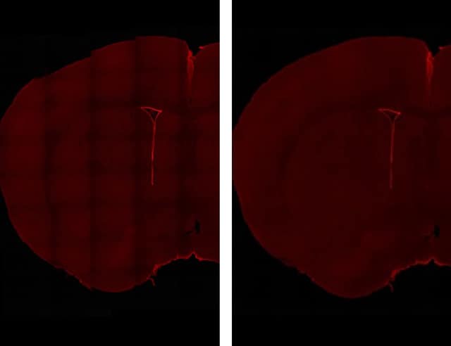
Stained images of ependyma cells of an adult mouse.
The image to the left is the raw data obtained by connecting together five horizontal images and eight vertical images that have been captured. The points of connection stand out because the contrast differs.
The image on the right, in comparison, is an image produced by the BZ Series image stitching function, which has automatically corrected the connection contrasts between the different images. These connections no longer stand out. From capturing the image to combining the images and correcting them takes under 3 minutes using the BZ Series.
(Photographs provided by: Assistant Professor Dr. Takehiko Sunabori, School of Medicine, Keio University)
Stained magnified images of ependyma cells of an adult mouse. The image on the left is a raw image with fluorescence blurring. The image on the right was obtained by eliminating fluorescence blurring using the BZ Series haze reduction function, thereby clearly revealing the target cells to observe. (Photographs provided by: Assistant Professor Dr. Takehiko Sunabori, School of Medicine, Keio University)
Also, the dynamic cell count function is useful for counting the number of cells in a sample. Thanks to its unique extraction method that separates individual cells on the basis of the change in brightness, instead of contour extraction through binary processing, it is possible to distinguish and count non-circular, severely adhered cells as well. This is said to have improved the accuracy of cell counting.
Together with functions helpful for research, the quick startup after power-up is also highly regarded among researchers. “Although conventional fluorescence microscopes require time and effort to make optimal settings, the BZ Series enables immediate observation any time we want. This user-friendliness is also well-valued among graduate students, who use the BZ Series often.”
Dr. Okano’s research facility stands at the global forefront of research into regenerative treatment of the nervous system. To remain at the forefront, however, this research facility must push through the rigorous international competition it faces. As research constantly advances, one of the greatest challenges is how to increase the efficiency of the observation of experiment samples. The BZ Series, with its beneficial functions and user-friendliness, provides strong support to the groundbreaking research conducted by the Okano Research Institute.
(As of July 2008)
Subsequent achievement by Dr. Hideyuki Okano
The world’s first successful treatment of spinal damage in mice using human iPS cells
The regenerative treatment of spinal damage that Dr. Hideyuki Okano engages in has advanced to a new stage. According to his announcement made at the 5th Keio University Advanced Science and Technology Symposium held on February 4, 2009, he succeeded in curing spinal damage in mice using human iPS cells for the first time in the world.
In this experiment, neural stem cells grown from human iPS cells were transplanted into mice with damaged spinal cords. As a result, most of the 29 transplanted mice showed significant recovery of motor functions compared to the non-transplant group to the extent that coordination was enabled between the fore and hind legs, with the mice placing weight on their hind legs.
As mouse nerve cells are structured similarly to those of humans, the success of this experiment can be said to be a big advancement toward human application. At present, treatment using iPS cells involves concerns about the side effect of tumor formation. Therefore, from the viewpoint of treatment safety, Dr. Okano intends to continuously pay careful attention to the progress of this experiment.
<General knowledge> Neural stem cells
Neural stem cells are stem cells that produce the neurons and glial cells that make up the nervous system. Conventionally, the common perception within the medical field was that adult mammalian brains did not produce neural cells. According to Dr. Okano’s research, however, it has been clarified that neural stem cells exist within the brain. This has led to a door opening on the path toward regenerative treatments of the central nervous system.
<General knowledge> iPS cells
iPS stands for “induced pluripotent stem cells.” These are pluripotent stem cells that are created from somatic cells. Theoretically, any cell composition within the body can be created from iPS cells. However, research is still in the very early stages, with many questions—including the issue of the tumorigenic transformation of cells during differentiation—still to be answered.

