Fluorescence Microscopes
The perfect solution for everyday patient diagnostics and clinical research at the Institute of Neuropathology in the Charité hospital in Berlin

Dr. Frank Heppner
Director at the Institute of Neuropathology
Charité – Universitätsmedizin Berlin
Dr. Frank Heppner has headed the Institute of Neuropathology at the Charité hospital in Berlin since 2007. With around 55 members of staff, it is the largest neuropathological institute in Germany and examines approximately 17,000 patient samples every year. The affiliated medical research department focuses on neuromuscular research, autoimmune diseases, neuro-oncology, brain tumors and lymphomas, childhood brain tumors and neurodegenerative diseases.
Since spring 2020, the institute has also conducted research on the effects of SARS-CoV-2 infection in the central nervous system (CNS). The first results of investigations into the pathway through which the virus can enter the brain were published in the Nature Neuroscience journal [1] in November.
Get detailed information on our products by downloading our catalog.
View Catalog

Logistical challenges must be easy to resolve
The Berlin Charité is one of the largest university hospitals in Europe and one of Germany’s top research institutes. Charité operates on the core facility principle, which means that large-scale equipment and expensive, highly sensitive measuring apparatuses are consolidated in central facilities to help ensure this technology is used effectively. As a result, not every university department has a full complement of equipment.
The research department and the routine laboratory of the neuropathological institute are located in two different buildings on the Charité Mitte campus. The scientists in the research department have an old fluorescence microscope, but it is often not capable of producing meaningful images. So they have to resort to an external confocal laser scanning microscope – and that requires time slots to be booked.
Previously, there was no fluorescence microscope in routine health care that could be used in medical diagnostics, so staff booked time slots on external microscopes as and when required. That meant Dr. Heppner could justify the acquisition of a fluorescence microscope for his institute to finally fill a gap that until now could only be bridged with careful, time-consuming planning.

A wide range of users with high expectations
Each individual specimen poses different challenges, and a new fluorescence microscope specifically for Dr. Heppner’s neuropathology department needs to handle all of them. From MTAs and master’s students to residents and researchers, there is a long list of criteria that needs to be met in terms of resolution, features, location, and most importantly, user friendliness.
The Institute of Neuropathology stipulated the following key criteria for a new fluorescence microscope:
- it should be practical, small and portable
- it must be compatible with the institute’s computers
- it needs an integrated darkroom, so that it can be used in any location
- it requires a high resolution comparable to that of a confocal laser scanning microscope for certain specimens
- it must be possible to eliminate fluorescence blurring
- it should have an integrated stitching function in order to merge multiple fields of view of a particular specimen
- it must be easy to integrate into the diagnostic workflow
- it must enable simple and complete documentation of patient samples
- it must be easy to use
As Dr. Heppner tells KEYENCE in an interview, if he had to design a fluorescence microscope for his institute, it would have all the features that the BZ fluorescence microscope offers. Whether it’s a quick view of the overall image or a detailed zoom into the individual cell, stitching or optical sectioning, simple operation or portability, the BZ fluorescence microscope has it all covered. So it’s practically tailor-made for him, effortlessly meeting the various requirements of neuropathological research and diagnostics.
The ideal location
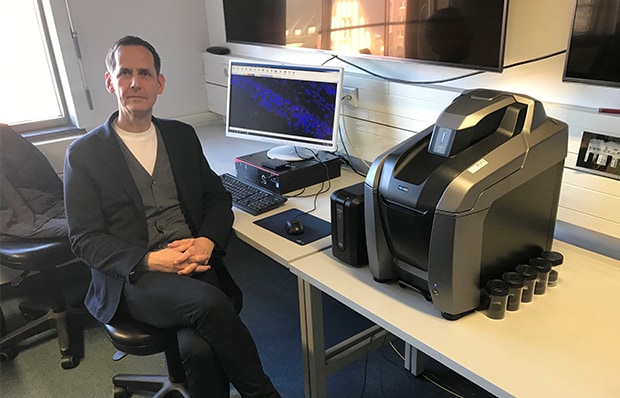
Figure 1 Dr. Frank Heppner in the microscopy consultation room with the BZ fluorescence microscope
Since research and patient care are spread over two buildings, the microscopy consultation room in routine diagnostics was chosen as the location for the microscope. With its integrated darkroom, the BZ fluorescence microscope can be set up in any location that offers high efficiency when conducting experiments. The microscopy consultation room is accessible to all staff at all times and features an impressive array of technology, including a network-compatible institute computer and a presentation monitor. The BZ fluorescence microscope connects to this hardware, allowing the fluorescence images to be used immediately for consultation and diagnostics as well as for light microscopic specimens. The fluorescence microscope was an instant success and the first images have already been incorporated into new manuscripts, Dr. Heppner tells us. He says it’s because the BZ is so easy to use: even inexperienced users can quickly generate meaningful, high-quality images.
Multiple specimens – multiple requirements
The large university neuropathology institute conducts routine diagnostics and has six research groups, which means various questions have to be answered using fluorescence microscopy. The variety of specimens ranges from human and murine tissue sections of the brain, brain tumors, cerebrospinal fluid samples, CNS and muscle tissue sections to cell cultures and single cells.
Challenges in routine diagnostics
In Dr. Heppner’s diagnostics laboratory, for example, FFPE sections are marked with LCOs (luminescent conjugated oligothiophenes) to detect amyloid substances when various muscle diseases are suspected. These don’t just form deposits in the brain, as in Alzheimer’s disease, but also in muscles and CNS nerves outside the brain. Fluorescence measurement is used to determine the biochemical configuration and conformation, which is critical for diagnosis. Another standard procedure in routine practice is detection of immunoglobulins in patients with autoimmune brain diseases using fluorescence microscopy. Previously, these investigations had to be conducted on research equipment in other buildings by booking a time slot.
For routine diagnostics, the advantages of purchasing the KEYENCE BZ fluorescence microscope are clear: the BZ is accessible to everyone at all times in the microscopy consultation room and is so easy to operate (thanks to its fully electronic control system) that no complicated, time-consuming training is required. Patient samples can immediately be analyzed and documented in full without the need for a scanner capable of fluorescence imaging. Another major advantage is that the doctors and researchers can now train on the job, making it easier for them to manage their time.
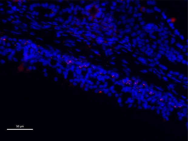
Figure 2 Colocalization of SARS-CoV spike protein in the olfactory mucosa of individuals with COVID-19
Fluid boundaries between research and diagnostics
Multiplex fluorescence has mainly been used in research at the Institute of Neuropathology, but it is now being used more and more in diagnostics. Colocalization of target structures in different cell types, especially as part of the research currently being conducted by Dr. Heppner’s research groups on COVID-19, offers an insight into how SARS-CoV-2 can spread from the olfactory mucosa to the brain. The translational results obtained by the Institute of Neuropathology and by more than 20 other participating research groups were published in Nature Neuroscience [1] at the end of November 2020. Epifluorescence microscopy was used to detect the spike protein of SARS-CoV-2 on neurons located in the olfactory mucosa, and the virus can be traced along the olfactory nerve into the brainstem. Multiplex fluorescence shows that the virus – at least so far – cannot be detected in neurons, suggesting that it can also enter the brain through the small blood vessels that run through the CNS.
However, even though the virus has not yet been detected in the brain, some COVID-19 patients exhibit late post-viral neurological problems even after acute symptoms have disappeared (known as long COVID). These are probably due to the reaction of the immune system to SARS-CoV-2, for example in terms of the cytokine storm of immune cells triggered in the brain. Research on long COVID is still in its infancy, and the CNS has an important role to play in these studies.
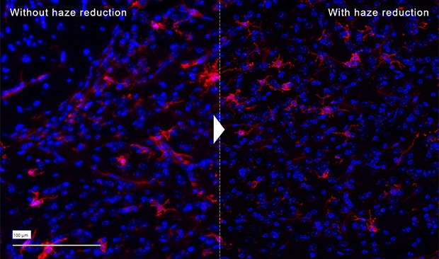
Figure 3 Image of microglia
Research challenges
Multiple sclerosis is a chronic inflammatory neurological autoimmune disease. Dr. Helena Radbruch’s research group at the Institute of Neuropathology is studying survival niches of certain immune cells in the brains of patients with this disease. The techniques used include sequential staining in the same section and spatially resolved transcriptome analyses. Both are very time-consuming and costly analyses, and the BZ fluorescence microscope has proved to be an efficient, resource-saving way to establish new stains for these techniques. Rapid imaging and sectioning make it easy to test in advance whether tissue samples are suitable for further analysis, and each antibody for individual brain structures such as glial cells, neurons, vessels, meninges, and various immune cell subtypes can be independently validated.
Over the last few years, Dr. Heppner has built up a biobank of autoptic brain material for Alzheimer’s research. The samples from this database usually show high autofluorescence caused by the degenerative deposits in the brain. This problem is part of everyday life for the research group, because Alzheimer’s sufferers often die at an advanced age. In the course of a lifetime, and especially in patients with degenerative brain diseases, many substances can be deposited that cause high autofluorescence. In these cases in particular, Dr. Heppner’s team is grateful that the BZ fluorescence microscope can remove fluorescence blurring in order to produce a clear view of individual cellular structures such as the autophagy apparatus or lysosomes.
Everyday research and routine work at the Institute of Neuropathology has been greatly simplified by the BZ fluorescence microscope, says Dr. Heppner. The fully electronic microscope generates fluorescence images quickly, and if problems occur they can even be fixed remotely by KEYENCE customer service. During a consultation, users no longer need to make the room awkwardly dark when showing fluorescence images. The BZ produces a high-quality resolution that meets the institute’s requirements; only in exceptional cases is an additional high-resolution confocal laser scanning microscope necessary. The stitching function is widely used in patient specimens and in slices of brain hemispheres from mouse models used in basic Alzheimer’s research. Finally, the elimination of fluorescence blurring is a major benefit because a large proportion of neurological specimens have autofluorescence, which produces lots of interference. In short, when it comes to microscopy, the Institute of Neuropathology at the Charité hospital in Berlin has a wide range of needs – and the BZ fluorescence microscope meets all of them.
[1] Meinhardt, J., Radke, J., Dittmayer, C. et al. Olfactory transmucosal SARS-CoV-2 invasion as a port of central nervous system entry in individuals with COVID-19. Nat Neurosci 24, 168-175 (2021). https://doi.org/10.1038/s41593-020-00758-5

