Mouse Cerebral Cortical Neuron Spine
Advanced observation delivers high-resolution images
A mouse cerebral cortex (DIV21) is immunostained with GFP antibody to observe it. This process makes it possible to observe the shape of the spine.
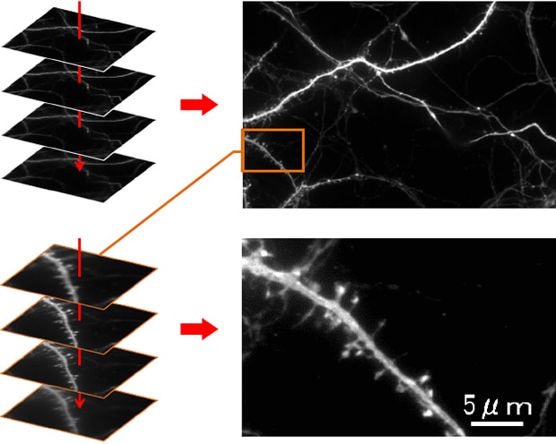
Conventional problems
With conventional microscopes, it is not possible to view images clearly due to noise after magnifying the details.
Also, thick samples like spines generate out-of-focus images, making it difficult to observe them clearly.
- Using the All-in-One Fluorescence Microscope BZ-X800
-
- The cooled monochrome camera enables low-noise observation with high sensitivity.
- Thanks to the camera’s high resolution, the clarity of the original image is not lost even after partial magnification or clipping, enabling you to observe even microscopic structures clearly.
- Here are some examples of using the All-in-One Fluorescence Microscope BZ-X800 in front-line research.
- [Myelodysplastic Syndromes (MDS)] Stitching, Sectioning and the Z-Stack Function as Decisive Arguments for the Acquisition of the BZ Fluorescence Microscope at the University Hospital of Düsseldorf
- [Neuropathology] The perfect solution for everyday patient diagnostics and clinical research at the Institute of Neuropathology in the Charité hospital in Berlin
- [Regenerative Medicine] BZ Series Provides Essential Imaging for Neural Stem Cell and Spinal Observation
- [Gene Therapy] Improving Research for the Development of Gene Therapy Drugs
- [Heart Disease Treatment] Developing Cell Sheets for Myocardial Regenerative Treatments
- [Cancer Treatment] Automated Fluorescence Microscope Transforms Process for Induced Cancer Stem Cell Research
- [Immune System] BZ Series Contributes to Understanding the Pathological Model of Asthma
- [Biomaterials] Promoting Efficiency in Research With Compact, User-friendly Microscopes