Fluorescence Microscopes
Localization Imaging of Exosomes
In recent years, there has been a focus on extracellular vesicles (EV), which are secreted by not only tumor cells and immune cells, but by all cells in the body. EVs are small particles surrounded by a membrane, typically between 30 - 150 nm in diameter, that are secreted by cells and exist in the majority of bodily fluids (such as blood, urine, and cerebrospinal fluid) and in cell cultures.
Extracellular vesicles are classified into exosomes, microvesicles (MV), and apoptotic bodies according to differences in the mechanisms with which they are generated. For a long time, exosomes were believed to participate in the ejection of unnecessary cell contents. However, in recent years, exosomes have been brought into the limelight because it has been confirmed that they have the potential to clarify the pathology of and to treat various diseases such as malignant diseases, immunologic diseases, neurological diseases, and infectious diseases.
This section introduces the basic knowledge of the generation mechanisms and functions of exosomes, examples of disease treatment, and an example of the observation of exosomes. We hope this information will serve as a reference for everyone who is interested in exosomes but is not sure how to observe and analyze them.
Get detailed information on our products by downloading our catalog.
View Catalog

What are exosomes?
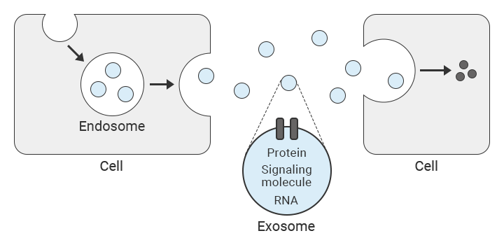
Exosomes are one type of extracellular vesicle. Cells bring extracellular materials into them via a mechanism known as endocytosis, during which vesicles known as endosomes are generated. Generated vesicles transition from clathrin-coated vesicles to early endosomes to late endosomes. Then, when a multivesicular body (MVB)—which contains many intraluminal membrane vesicles (ILV)—fuses with the cellular membrane, membrane vesicles are ejected from the endosome out of the cell. These ejected endosomes are exosomes.
Exosomes contain various proteins and ribonucleic acids (RNA) derived from the secreting cell such as proteins derived from endosomes and the cellular membrane and proteins related to intracellular transport. They also contain the cellular membrane of the secreting cell and fats derived from the endosome membrane. These contents are known as the cargo. These exosomes are then taken in by other cells, at which point, the proteins and RNA contained within the exosome are transmitted to the new cell.
From this series of actions, it is believed that exosomes have a key role in intercellular communication. Hence, research into exosomes is ongoing, especially in the medical field.

Uptake and exosome functions
Although the exosome uptake mechanism used by cells is not clearly defined, the need for recipient cells to recognize molecules on the surface of an exosome has been identified.
The outer layer of an exosome is composed of lipid rafts, membrane transport proteins, and transmembrane proteins such as CD9, CD63, and CD81. The lipid composition also varies according to the origin cell. Uptake also depends on whether the receptor cell is composed of cells with molecules able to recognize the exosome’s surface molecules.
In this way, the molecular makeup of the exosome’s outer layer and the receptor cell itself are considered important for the in vivo dynamics of the exosome.
After an exosome is taken up by a cell, the exosome transmits information to the receptor cell using nucleic acids. The receptor cell then synthesizes protein based on the information in those nucleic acids. Understanding of this led to the discovery that exosomes secreted by immune system cells can suppress the expression of certain genes and promote immune responses.
For example, exosomes secreted by dendritic cells—a type of antigen-presenting cell—contain MHC molecules that can be used to activate T cells not located near dendritic cells.
Treating diseases with exosomes
The clarification of the function of exosomes has led to an increase in the activity of research into treatment and diagnostic methods that apply this function for internal secretion and circulatory organs. Examples include the early detection of cancer and treatment of neurodegenerative diseases such as Alzheimer’s dementia, Parkinson’s disease, spinocerebellar degeneration, Huntington’s disease, and amyotrophic lateral sclerosis (ALS).
Early detection of cancer
It is believed that exosomes participate in factors such as the existence, malignant progression, and spread of cancer cells. The exosomes ejected by the cancer cells of cancer patients contain various proteins derived from the cancer cells and bring about the activation of cytotoxic T cells characteristic of cancer cells. The reason is because exosomes derived from cancer cells contain cancer genes. Consequently, observation of the exosomes ejected by cells in cancer patients can lead to the early detection of cancer.
Regarding cancer diagnostic methods in which the focus is on the property of exosomes of conveying information from one cell to another, early detection of cancer is possible by way of liquid biopsy. In cancer diagnosis by way of liquid biopsy, it is believed that early detection of cancer is possible through an investigation of the exosomes of abnormal cells because the exosome contents show the characteristics of the cells that eject (circulating tumor DNA [ctDNA] that has variations to cancer-related genes).
Treatment of Alzheimer’s dementia
Alzheimer’s dementia is a type of cognitive impairment caused by the brain atrophying due to nerve cells dying because of damage caused by the toxicity of amyloid beta. In the treatment of Alzheimer’s dementia as well, it is believed that endosomes have a major effect on the increase or decrease of amyloid beta and tau protein,*4 which are the substances that cause the disease.
Also, there are ongoing attempts at techniques to enclose items such as medicine and nucleic acid in exosomes in a drug delivery system (DDS) that efficiently delivers the medicine to the specific diseased tissue and cells in a safe manner.
*4: Tau protein
Tau protein exists in the nerve cells of the central nervous system and peripheral nervous system. It bonds with various proteins and is concerned with various phenomena that occur in the central nervous system such as the maturation of the post-natal brain and the neural development of adult organisms. It is believed that abnormalities in tau protein cause neurodegenerative diseases such as Alzheimer’s dementia.
Regenerative medicine applications
Regenerative medicine based on exosomes can be used to regenerate and recover damaged organs and tissues by transplanting tissues grown from stem cells and other materials into a patient’s body.
Exosomes secreted by mesenchymal stem cells (MSCs) have therapeutic effects on various diseases such as cancer, renal disease, myocardial disorders, brain disease, and lung disease. MSCs can also be differentiated into endodermal and ectodermal lineage cells, and molecules secreted by undifferentiated MSCs have been shown to have a therapeutic effect against numerous non-specific diseases.
MSCs are also able to spontaneously search for and concentrate on damaged sites, referred to as homing. This ability is believed to make administering medicine intravenously to organs and tissues possible. The MSCs that accumulate in a damaged area then secrete MSC exosomes that regenerate and restore the tissue.
In this way, regenerative medicine based on MSC exosomes incorporates a different approach to cell therapy than with iPS cells, and this approach is attracting attention particularly for treatment of new virus infections.
Diagnosis with liquid biopsy
Liquid biopsy is a technique with a minimal effect on the human body. This technique uses body fluids (minimally invasive liquid samples such as blood and urine) and is useful mostly in cancer diagnosis.
Liquid biopsy samples contain tumors, various tissues, and cell-derived information. Observation of such samples can give a bird’s-eye view of the tumor profile of the entire body.
With the biopsy technique, in which a sample is harvested with a conventional endoscope or needle, regardless of subjecting the patient to pain and risk of complications, it is only possible to harvest a small portion of the tumor tissue. As such, it was only possible to detect fragmentary profile information with this technique.
With a liquid biopsy, inspection is possible with just body fluid, so cancer gene diagnoses and predictions of treatment effectiveness can be performed with no effect on the body.
What is a drug delivery system (DDS)?
A drug delivery system is a treatment method that uses the property of exosomes of transporting materials (proteins, nucleic acid, and fats) from one cell to a specific cell.
In the past, there have been examples of anticancer effects being confirmed in breast cancer cells and the amount of amyloid beta—a substance that causes Alzheimer’s dementia—contained in neuroblastoma cells being reduced with this treatment method.
Because exosomes are biomolecules, it is said that treatment by way of a DDS is less toxic than the conventional medicine transportation method using nanoparticles. Also, because it has been confirmed that a DDS can transmit medicine to tissues that were difficult to reach using conventional techniques, there are high hopes for this method as a powerful transportation molecule.
Future potential of exosome research
As outlined above, because exosomes convey information between cells, they have the negative characteristic of also transmitting disease. However, as medical science and observation technology develop, this characteristic is the very factor that is leading to people focusing on exosomes as a secret weapon that can open up the advanced treatment of prevention, early detection, and direct administration of medicine to cells.
Furthermore, exosomes are also secreted from the cells of vegetables and fruits. For example, ginger has the effect of suppressing alcohol-induced liver damage, but this is believed to be because the exosomes derived from ginger protect the liver from damage. There have also been reports of research results showing that the exosomes of boiled eggs suppress the hardening of the arteries and improve memory.
In Eastern medicine, diet is the root of medical treatment, and there is the belief that, essentially, the root of medicine that heals sickness and diet that promotes health is the same. From this point of view, by opening up a wide range of possibilities for both medical treatment and food, exosome research can be said to be the modern version of the concept that a balanced diet leads to a healthy body.
Exosome detection
There are two main types of exosome analysis methods. One method is to extract exosomes from body fluids or cell cultures and to analyze the proteins, fats, and RNA transported by the exosomes. The other method is to directly detect and analyze exosomes from body fluids. Furthermore, observation and analysis using a fluorescence microscope are performed in the tracing of items being taken into and secreted from cells.
Method using body fluids or cell cultures
Generally, techniques such as ultracentrifugation and ultrafiltration are used to separate and purify exosomes from samples of body fluids such as blood and ascitic fluid. However, it is difficult to analyze only the exosomes with these techniques because the sample also contains other items such as particles with similar characteristics and concentrations and high-weight proteins.
Hence, this method is sometimes performed in combination with affinity purification.*1 Also, size confirmation and western blotting*2 are performed with dynamic light scattering and a microscope in the final stage to check that the separated and purified sample is indeed a group of exosomes.
*1: Affinity purification
This is a technique for separating and purifying the target molecule, protein, or complex body through use of the reaction between the target molecule and a molecule (ligand) that bonds with it in a specific and reversible manner.
*2: Western blotting (WB)
This is a basic experimental technique for determining the characteristics of proteins. This technique is suited to measuring proteins that appear as exosomes.
Direct detection from bodily fluids
The techniques of flow cytometry,*3 microarray analysis, and surface plasmon resonance are being researched as ways to detect exosomes from body fluids without having to separate or purify the exosomes. Another technique, known as ExoScreen, developed for the diagnosing of large bowel cancer is also available. These four techniques are mainly used in the treatment of cancer.
In place of biopsy (in which a conventional endoscope or needle is used to harvest tumor tissue), there are high expectations for liquid biopsy (in which diagnoses and predictions of treatment effectiveness are performed on a sample of blood or other such body fluid) in terms of early detection of illnesses.
*3: Flow cytometry
This is a technique for evaluating particle characteristics by using the scattered light or fluorescent light obtained when a cell or minute particle is irradiated with a laser beam. It can be used to measure at high speed multiple characteristics of a single cell (cell, bacteria, etc.) at the same time. It is suited to the evaluation of the level of expression of exosome proteins and the evaluation of the taking in of exosomes into cells. It is difficult to classify fixed quantities and the vesicle type, but this method is the easiest way to observe the behavior with which exosomes are taken into cells.
Exosome observation and analysis
Exosomes have diameters of 30 to 150 nm, so they are difficult to observe with optical microscopes. Also, electron microscopes pose the problems of troublesome sample preparation and sample deterioration due to the vacuum and electron beam. In addition, electron microscopes are not commonly used to measure physical properties. Most importantly, electron microscopes are not available in every facility.
With exosome observation using a fluorescence microscope, it is possible to visualize and trace exosomes by way of fluorescent labeling. The RNA contained in the extracted exosome (the RNA cargo) or the exosome membrane is visualized by way of staining with a fluorescent dye and is thereby observed.
This observation enables the monitoring of time-dependent exosome localization changes and of exosomes being taken into cells (a process of intercellular communication via exosomes). Fluorescence microscopes can be used to perform observations, such as of exosome changes and the corresponding process, that are not possible with other methods. Therefore, fluorescence microscopes are receiving attention as a new piece of technology that enables analysis with higher accuracy.
Observation of 3D localization of exosomes using a fluorescence microscope
Localization is difficult to observe with normal fluorescence microscopes because it is hard to bring the image into focus. Also, flow cytometers, which evaluate particle characteristics by using the scattered light or fluorescent light obtained when a cell or minute particle is irradiated with a laser beam, are not able to check localization or evaluate changes over time.
The unique optical sectioning technology in the All-in-One Fluorescence Microscope BZ-X uses an electronic projection element for structured illumination to realize hitherto unseen clarity in images. Localization and changes over time are both displayed clearly.
In the following example, the sectioning function of the BZ-X is used to observe the localization of exosomes within a human epidermal cell.
With the BZ-X, it is possible to use high-resolution images to observe the movement of exosomes generated by a cell into other cells.
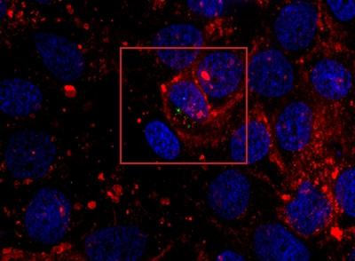
Green: exosome, red: cell membrane
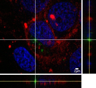
XYZ slicing image capturing
Objective lens: Plan Apo 100x
Exosome: green (PKH67), cell membrane: red (CellMaskTM Orange), cell nucleus: blue (DAPI)
Using the All-in-One Fluorescence Microscope BZ-X
- Fully motorized operation makes it easy to navigate around the sample and improves imaging efficiency.
- Capture clear images without the influence from out-of-focus fluorescence, even on thick or complex tissues, with the optical sectioning function.
- Evaluate cellular structure and localization by combining high-resolution image slices into a 3D composite.
- Cultivate and easily analyze cells to monitor changes over time.

