Fluorescence Microscopes
There are a number of objectives when observing hair, including assessing damage from perms or dyeing, or the penetration of hair care products. In particular, the cuticle is most susceptible to such processing, so observing the cross-section of hair in high definition is essential to evaluation. Here, we explain the structure and components of hair, the issues with conventional observation methods, and how they can be solved.
Get detailed information on our products by downloading our catalog.
View Catalog

Hair structure
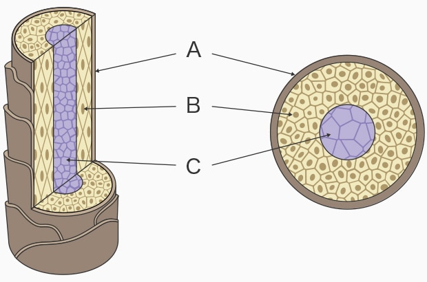
A: Cuticle B: Cortex C: Medulla
Hair is made of keratin, which is keratinized protein. It corresponds to the stratum corneum, or horny layer, and dead skin cells on the skin. The shaft, which is the visible part of hair, is made up completely of dead cells, which means that once they are damaged, they do not regenerate.
The hair shaft consists of three layers, from outermost to innermost: cuticle, cortex, and medulla.
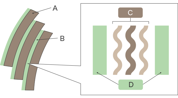
A: Cuticle B: CMC C: Delta layer (protein) D: Beta layer (CMC lipids)
Cuticle
The cuticle is composed of overlapping cells that cover the surface of the hair shaft like scales. One cuticle cell is 0.5 to 1 μm thick, clear and colorless, with moisture-repellent properties. Cuticle cells are aligned in the same direction—you can smoothly run your fingers through your hair from the root, but may feel some scratchy resistance if you try doing this in reverse.
One cuticle cell covers about 1/3 to 1/2 of the circumference of the hair shaft. If you have stiff hair, you may have more cuticle cells covering the circumference. The gaps between the cuticle cells are filled with cell membrane complex (CMC).
The CMC is composed of beta layers (lipids) that sandwich a delta layer (protein). The thickness is 0.04 to 0.06 μm. Hair dye and other hair products penetrate the hair through the CMC.
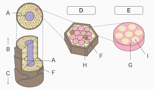
A: Cortex B: Tip of hair C: Root of hair D: Cortical cell E: Macrofibril F Melanin G: Matrix H: Amorphous keratin I: Microfibril
Cortex
The cortex is a cluster of macrofibrils. Each macrofibril is an assembly of microfibrils, and the gaps between them are filled with a hydrophilic matrix with hydrating properties. The main component of this matrix is amorphous keratin. It contains a rich amount of melanin pigment, along with amino acids, polypeptides (PPT), nucleic acid, and minerals. The matrix is the part affected by perms or dyed by hair dye, so it is important for hair care.
Medulla
The medulla is at the center of the hair shaft and is made up of elongated cubic cells made of protein and lipids. It contains air between the cells, and this gap reflects light. When this gap enlarges, the hair appears whiter.
Not all hair has medulla. Thin hair and fine hair do not have medulla. It may also be discontinuous in some spots.

High-resolution observation of hair
Optical microscopes or scanning electron microscopes (SEM) are used for high-resolution observation of hair.
Microscopic observation is done under specular reflection light by observing hair from the side using coaxial illumination. Observation of flat surfaces under specular reflection light is bright due to the reflected light directly entering the lens or CCD. In contrast, slopes in stepped parts appear darker due to some reflected light escaping off the surface. This contrast allows for fine structural visualization of the surface of hair. However, the surface of hair is curved, and reflected light escapes outward even more on the edges of the hair. Cross-section evaluation using an optical microscope does not provide the resolution necessary to observe the laminar structure of the cuticle, which may result in images of a single cuticle layer.
On the other hand, while a scanning electron microscope (SEM) allows for high-resolution observation of the cuticle surface structure, observation of non-conductive specimens requires pretreatment to prevent charge-ups, in which the incident electron beam accumulates on the surface of the specimen and creates a foggy glow. This can take a lot of time and effort. In addition, the hair must be placed inside a vacuum chamber for observation under vacuum, which causes the moisture in the hair to evaporate and may change the hair’s morphology. Whichever approach is used, it may be difficult to observe hair in its natural state.
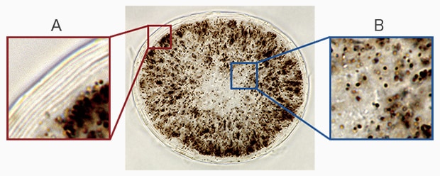
A: Laminar structure of cuticle B: Melanin found across the cortex
With the high-resolution offered by our BZ-X All-in-One Fluorescence Microscope, you can clearly observe specimens with laminar structures like the cuticle.You can also observe the melanin spread across the cortex in clear view, and simultaneously evaluate the cortex and cuticle.
Sample: Cross-section of human hair
Imaging magnification: Objective lens 100x (oil immersion)
Imaging method: Brightfield
Using the All-in-One Fluorescence Microscope BZ-X
- Using the BZ-X allows for both full cuticle observation and fluorescence microscopy in normal observation mode.
- Through high-resolution, cross-section observation, you can simultaneously evaluate the laminar structure of the cuticle and the melanin in the cortex.
- Combined with fluorescent labeling, you can also observe the penetration of hair care products into the hair. The BZ-X’s ability to quantitatively analyze multiple samples supports quantified evaluation.

