Fluorescence Microscopes
Image stitching & quantification
Automatic stitching
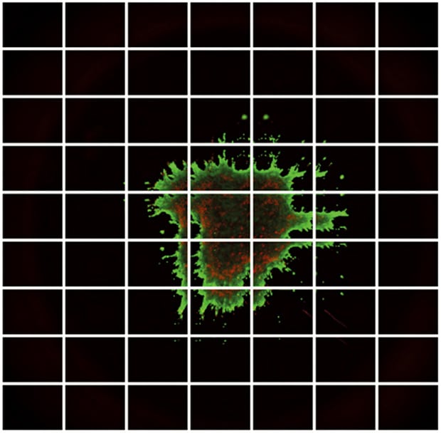
The growth potential of cells cultured under different conditions can be evaluated by calculating the ratio of live cells to dead cells. In the following images, fluorescence microscopy is used to differentiate the two: live cells are fluorescing green, whereas dead cells are fluorescing red. For accurate quantification, observation needs to be performed at a magnification that enables the shape of each cell to be captured clearly.
However, the high magnification required limits the amount of the cell that can be seen, and maintaining focus becomes more difficult as cells grow thicker in three-dimensional culture. This complicates image capture, which in turn introduces variability to the data output.
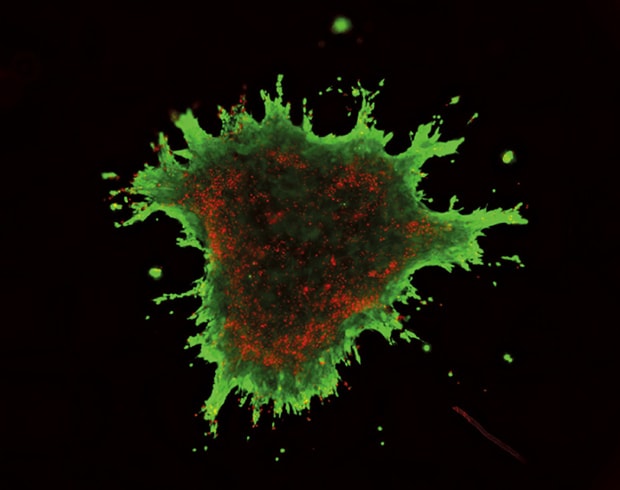
With integrin (cell adhesion molecule)
Live cell area ratio: 67.2%
Without integrin
Live cell area ratio: 73.3%
Using the All-in-One Fluorescence Microscope BZ-X
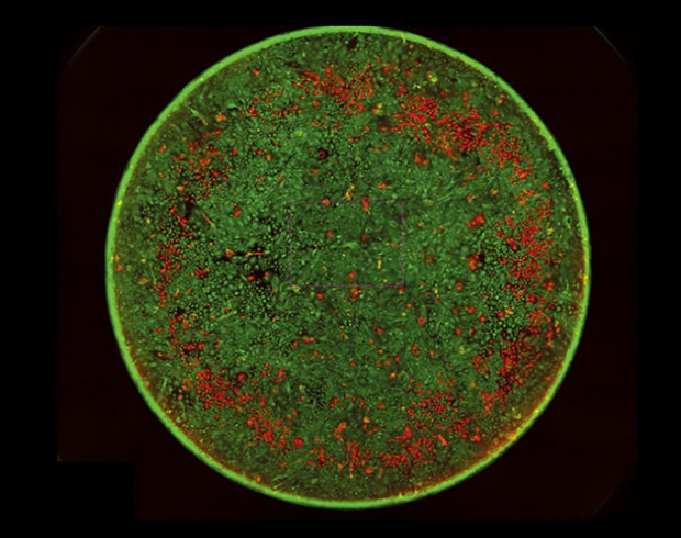
- If the entire specimen cannot be captured within one field of view, simply specify the desired outer bounds for image capture. The stage will automatically adjust to capture images across the specified area. Captured images can be stitched automatically to provide a single, high-resolution, wide-field image.
- Even for thick sections or samples with height variance, multiple images can be captured in the Z direction to focus on structures in multiple focal planes. This produces a fully focused, clear image of the entire sample.
- Quantitative measurement can also be performed on stitched images with Hybrid Cell Count, enabling high-precision analysis of the entire sample.


