Fluorescence Microscopes
Cell Cycle Observation of an Oral-derived Epithelial Cell Line (Ca 9-22) Using FUCCI
Example of cell cycle observation using time-lapse imaging
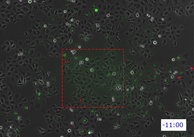
FUCCI (Fluorescent Ubiquitination-based Cell Cycle Indicator) is a fluorescent probe that is effective in monitoring the progression of a cell cycle in real time. The video shown below has been captured using KEYENCE's All-in-one Fluorescence Microscope BZ-X to perform time-lapse imaging of an oral-derived epithelial cell line (Ca 9-22) in which FUCCI has been introduced. The imaging was carried out over a total of 60 hours at an interval of 30 minutes (for a total of 120 images).
By using the Multi-point Image Capturing function, 3 points per well of a 6-well plate (for a total of 18 points) were captured continuously, once every 30 minutes.
It is also possible to zoom in on a specific part.
Provided by:
Yoshikazu Mikami
Niigata University, School of Medicine
Basic Medicine System, School of Microscopic Anatomy (Third School of Anatomy)
Get detailed information on our products by downloading our catalog.
View Catalog

Problems in cell cycle observation
To perform long-term time-lapse imaging using live cells, it is important to maintain constant temperature, CO2 concentration, and humidity. One simple method is to take the specimen out of the incubator only when capturing the image. However, this method poses several issues, including: difficulty in reproducing the same imaging location, possibility of contaminating or disturbing the specimen, and just the need for a user to have to manually move the sample each time.
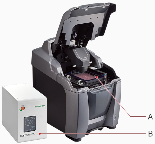
A:Stage-top incubator B:Temperature and CO2 controller
The All-in-One Fluorescence Microscope BZ-X uses a compact, stage-top incubator, thereby making it possible to automatically capture the specified number of images while still maintaining a constant specimen environment. Furthermore, it is possible to capture images in multiple locations while changing the magnification, filter and other imaging conditions automatically. This makes it possible to not only easily perform time-lapse imaging, but also to carry out experiments far more efficiently than with conventional methods.
Using the All-in-One Fluorescence Microscope BZ-X
- Fully motorized for easy operation. Because no darkroom is required and a compact incubator can be installed in the stage, the BZ-X can be used to easily start live cell imaging.
- A highly sensitive optical system composed of a cooled camera and a dedicated lens makes it possible to obtain high-resolution fluorescence images with minimal cell damage.
- Multiple samples can be time-lapse imaged at the same time, making it easy to perform comparative verification under the same conditions.

What is FUCCI?
This is technology that visualizes the progression of a cell cycle and makes it possible to observe the DNA reproduction and cell division status in real time by associating the fluorescent protein mKO2 (monomeric Kusabira-Orange2), which emits red fluorescent light, to Cdt1—which undergoes the greatest amount of growth in the G1 period in which a cell that has completed division prepares to reproduce DNA—and by associating the fluorescent protein mAG (monomeric Azami-Green1), which emits green fluorescent light, to Geminin—which undergoes the greatest amount of growth in the S, G2, and M periods that cover from DNA reproduction to cell division.
Since its development in 2008, FUCCI has been applied widely to furthering the understanding of the phenomena of life related to cell cycles in embryology, regenerative medicine, cancer oncology, and similar fields and is continuously being improved with examples including FUCCI (N+C) in which not only the interior of the nucleus but also the whole cell is labeled with FUCCI signals; FUCCI2 that makes the G1, S, G2, and M periods easier to separate by using mCherry and mVenus with the fluorescent proteins; and FUCCI (CA) that can distinguish all four cell cycles by adding a capability to distinguish cell cycle interphases (the G1, S, and G2 periods).

