Fluorescence Microscopes
What are the islets of Langerhans?
The islets of Langerhans, also called the islands of Langerhans, are the endocrine part of the pancreas.
The pancreas has exocrine cells that secrete amylase and other digestive enzymes into the duodenum and endocrine cells that secrete hormones. The exocrine cells make up most of the pancreas. The endocrine cells exist sporadically as small clusters like islands among the exocrine cells. These clusters were named the “islets of Langerhans” after German pathologist Paul Langerhans, who discovered them.
Insulin and other hormones secreted from the islets of Langerhans adjust blood glucose levels, and a weakening of this function is understood to cause diabetes.
Get detailed information on our products by downloading our catalog.
View Catalog

Islet cells and their functions
The pancreas can be divided into the head, body, and tail parts; the islets of Langerhans are mostly distributed in the tail of the pancreas.
The islet cell types, alpha, beta, and delta cells, each secrete different hormones, and regulate the concentration of glucose in the bloodstream. This is what maintains a normal blood glucose level.
α cell (A cell)
The α cells secrete a hormone called glucagon, which acts to raise the blood glucose level. When the blood glucose level drops, glucagon induces the breakdown of glycogen to raise the concentration of glucose in the bloodstream. At the same time, glucagon induces the release of glucose stored in cells into the bloodstream. With these two functions, glucagon raises the blood sugar level by raising glucose concentration.
β cell (B cell)
The β cells secrete a hormone called insulin. Insulin acts to lower the blood glucose level. When the blood glucose level rises, insulin induces the synthesis of glucose to lower the concentration of glucose in the bloodstream. Glucose synthesized by the working of insulin becomes glycogen, which is stored as energy, thus suppressing the release of glucose into the blood. At the same time, insulin induces the cells’ intake of glucose. With these two functions, insulin lowers the blood sugar level by reducing glucose concentration.
δ cell (D cell)
The δ cells secrete a hormone called somatostatin. Somatostatin is secreted when you take in food. In addition to suppressing the secretion of glucagon and insulin, it also inhibits the secretion of hormones that induce pancreatic secretions, such as cholecystokinin and secretin, from the duodenum. The inhibition of pancreatic secretion by somatostatin helps keep blood sugar levels within the normal range. At the same time, it can suppress excessive motility of the digestive system as well as absorption and consumption of nutrients.

Observing the islets of Langerhans
Fluorescent immunostaining using anti-glucagon antibodies and anti-insulin antibodies allows for visualization of glucagon-positive cells as α cells and insulin-positive cells as β cells, clarifying each cell type’s distribution in the islets of Langerhans. Use of a cell count application also makes it possible to quantitatively evaluate the positive cells’ area and fluorescence intensity. Research into transplanting pancreatic cells produced from iPS cells and other human pluripotent stem cells is underway as a recent radical therapy for diabetes, and in such studies it has been reported that the proportion of the area of insulin-positive cells is high in a diabetic mouse-derived graft. A quantitative method using fluorescent immunostaining is used for such evaluation.
Normally, an objective lens with a magnification of 4x to 20x is required for such evaluations, but since the whole specimen does not fit in a single field of view, researchers observe and capture images with a focus on the parts with islets of Langerhans. However, high demand for objectivity of evaluation data in recent years, along with the issue of eliminating arbitrariness* from imaging and analysis, means that individual observation and imaging of specific parts is incapable of meeting current needs.
The BZ-X All-in-One Fluorescence Microscope offers an image stitching function, where the operator can simply specify four points on the outer perimeter of the specimen to have the system automatically capture images and join them together with high precision. This function can produce a high-definition image of entire specimens, providing a solution to the previously mentioned issue.
In the following example, a 3 by 5 grid of images is automatically captured using the Plan Apochromat 4X lens and stitched together with the image stitching function to fit the pancreatic specimen into a single image.
* Eliminating arbitrariness:
Arbitrary means that there is no logical necessity for criteria and that decisions are made at random on the spot. Eliminating arbitrariness means to eradicate such arbitrary properties. In scholarly papers, great importance is placed on objectivity and reproducibility. Images captured with microscopes can make major contributions to these two goals. When partial images are captured of sections, the person capturing the images may sometimes do so in an arbitrary manner to capture a favorable location. The BZ-X captures seamless, high-resolution images of the entire specimen, eliminating arbitrariness attributable to the person capturing the images and, consequently, making it possible to perform more objective evaluations.
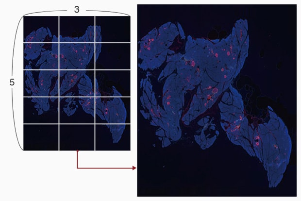
Objective lens: Plan Apochromat 4X
Image stitching: 5 images x 3 images
Using the All-in-One Fluorescence Microscope BZ-X
- When a slice of the target is too large to fall within a single field of view, images are captured while moving the stage and a high-resolution image can be created by stitching these images.
- Even for tilted specimens or specimens that have height differences, it is possible to create an image in which the entire specimen is in focus. This is accomplished by capturing multiple images in the Z direction and stitching together only the parts that are in focus.
- The Haze Reduction function, which eliminates fluorescence blurring caused by scattered light, can be used to capture clear images with high contrast.

