Fluorescence Microscopes
High-resolution Imaging of the Periodontal Membrane of Mice
Capturing clear images without fluorescence blurring
The periodontal membrane, also known as periodontal ligament, is a connective tissue that attaches the tooth root to the alveolar bone (upper and lower jawbones).
This fibrous tissue also adjusts bite forces applied to the tooth (impact absorption).
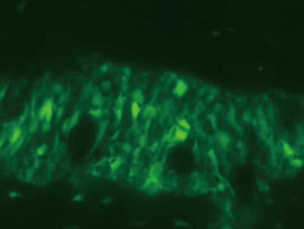
Normal observation
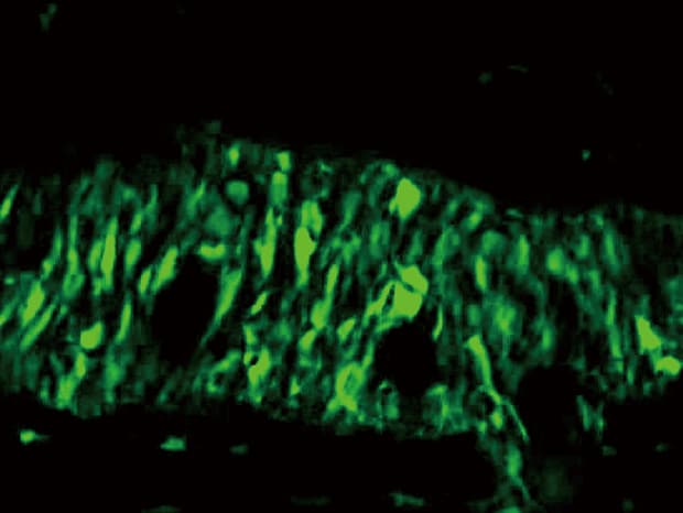
Sectioning observation
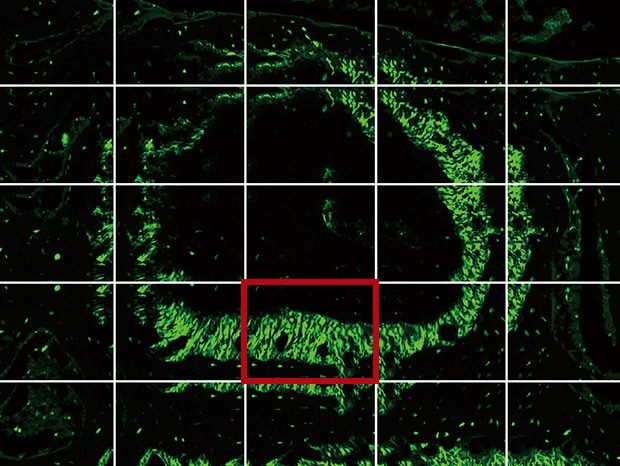
Image stitching
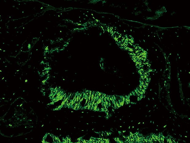
Objective lens: CFI Plan Apo λ 40x
Image stitching: 5 images × 5 images
Using the All-in-One Fluorescence Microscope BZ-X
- The Sectioning function makes it possible to eliminate fluorescence blurring optically and capture clear images.
- A built-in Z-stack function captures multiple images at different focal positions and is able to create a fully focused image by combining only the areas that are at their sharpest focus.
- When a slice of the target is too large to fall within a single field of view, images are captured while moving the stage and a high-resolution image can be created by stitching these images.
- Each cell can be extracted and counted using Hybrid Cell Count.

Image stitching
Up to 50,000 × 50,000 pixels can be joined together easily to capture a clear image without stitch lines or brightness variations. Additionally, it is possible to accurately quantify information such as fluorescent signal intensity.