Fluorescence Microscopes
Capturing clear images without fluorescence blurring
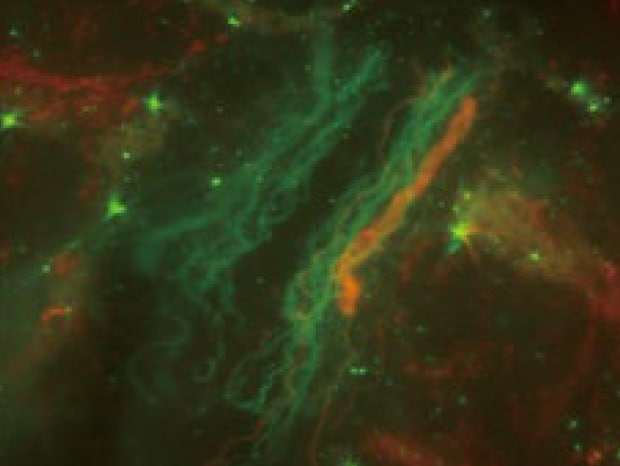
Zebrafish are used for various types of research because live tissues and cells can be observed due to their transparent bodies. However, determining localization can be difficult since spinal cords and neurons extend three-dimensionally, making them susceptible to fluorescence blurring when imaged under a conventional fluorescence microscope.
Normal observation
Sectioning observation
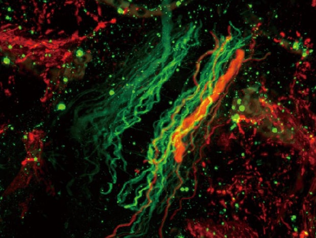
Objective lens: CFI Plan Apo λ 100xH
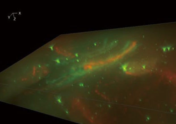
Sectioning + Z-stack
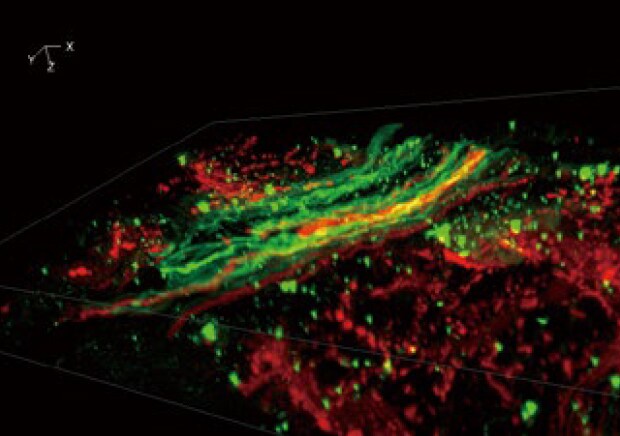
3D image construction
After images are captured at different focal planes, these images can be combined and displayed in 3D with a single click. Users can then rotate, zoom in or out, and analyze cross-sections of the 3D image to determine the fluorescent signal localization.
Normal observation
Sectioning observation
Using the All-in-One Fluorescence Microscope BZ-X
- The Sectioning function makes it possible to eliminate fluorescence blurring optically and capture clear images.
- A built-in Z-stack function captures multiple images at different focal positions and is able to create a fully focused image by combining only the areas that are at their sharpest focus.
- High-resolution 3D images can be created by combining a Z-stack of 2D images, allowing for clear observation of localization.


