Fluorescence Microscopes
Golgi staining
Golgi staining is a silver staining technique that visualizes dendritic spines by staining the axons and dendrites of fine brain nerve cells in black, which has long been used to observe the structure and changes of nerve cells in rat and mouse brain specimens.
Staining using Golgi’s method was discovered in 1873 by Camillo Golgi, an Italian Nobel Prize winner for Physiology or Medicine, who was working on experiments of metal impregnation of nerve tissue (silver staining). This method used to be called black reaction because when a brain specimen fixed with a fixative solution containing osmium tetroxide and potassium dichromate was stained in a silver nitrate solution, the fine tissue of the nerve cells turned black. Golgi-Cox staining, discovered by a Dutch doctor, Cox in 1891, uses improved compounds. A brain section fixed with a mixture of potassium dichromate, potassium chromate, and mercury chloride solution turns black when immersed in an ammonia solution, which provides more stable results.
Golgi staining and Golgi-Cox staining are carried out according to their respective protocols. To achieve higher work efficiency, including shorter staining times, and higher stability for specimen creation, various improvements have been made in the reagent. As a result, both tissue staining methods are still used for the observation of fine nerve tissue stained in brain sections. These methods stain only certain nerve tissue in a brain section, which makes them useful for research on various topics, such as determining how the shape and change of dendrites in the hippocampus affect cognition and memory formation.
Get detailed information on our products by downloading our catalog.
View Catalog

High-resolution observation of Golgi-stained brain sections
A brain achieves higher functions, such as memory, using networks formed with an axon extending from each nerve cell and a vast number of other nerve cells connected to each other via synapses. To observe brains during experiments and research, several specimens are created from an animal brain. A brain is sliced into pieces of the same thickness using a cryostat at low temperatures and stained according to the Golgi staining protocols.
Rat and mouse brains used as specimens are very small, and thus have extremely fine axons and dendrites in their nerve cells, which need to be observed at high magnifications using a microscope. It is, however, difficult to focus exactly on entire fine nerve cells in a brain section because the section has a certain thickness. Even a slight tilt or height difference in a specimen greatly affects focusing. One challenging issue is to obtain images that are clear enough to be used for thesis presentations and conferences, as well as high magnification observations and evaluations. In high magnification observations, it is important to acquire clear images of nerve cells. At the same time, it is also important to always understand which part of the brain section is being observed. As magnification is increased to achieve the necessary resolution, the field of view becomes narrower. To understand the entire brain section, it is necessary to observe a vast number of fields of view. To perform high resolution imaging over a large area, multiple images must be captured at high-magnification and stitched together. However, it is cumbersome and difficult to record and manage numerous high magnification images and stitch them manually, which requires a lot of time and effort.
The solution: High-resolution observation of a brain section
Operators need a high level of skill to manipulate a microscope in observation and to create specimens by staining brain sections. Even experts need to focus and expend a great deal of energy.
KEYENCE has fully motorized the drive system including focusing and stage movement, simplifying various cumbersome and advanced processes required in microscope observation so they can be carried out using only a mouse. KEYENCE has developed the All-in-One Fluorescence Microscope BZ-X that can both capture clear images at high magnification and record vast amounts of data, allowing users to quickly analyze entire specimens.
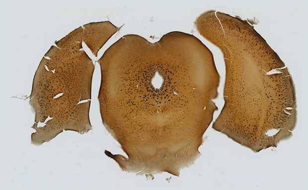
Seamless high-resolution observation of entire images and high magnification images
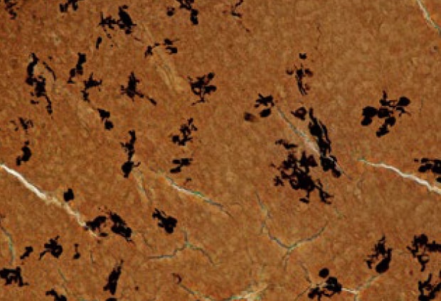
The following observation images are a Golgi-stained rat brain section captured using the BZ-X.
If a specimen is tilted, thick, or has height differences, conventional systems are only able to bring a portion of the specimen into focus, making it difficult to understand nerve cells in 3D. The BZ-X uses Z-stack to automatically capture multiple images in the Z-axis direction and the full-focus function to compose only the parts of each image that are in focus, allowing for easy capture of fully focused images. This makes it possible to observe fine nerve cells and Golgi-stained black sections clearly.
Additionally, the BZ-X is equipped with image stitching that captures multiple high resolution images at high magnification while automatically moving the motorized stage in the X- and Y-axis directions. This makes it possible to capture a high-resolution fully focused image of the entire brain section that is too large to fall within a single field of view when observed at high magnification.
The full focus function and image stitching can be used together, which allows users to observe fine nerve cells at high magnification while always understanding which part of the brain section is being observed.
Image construction using full focus and image stitching
A Z-stack is performed to capture multiple images over a single field of view at varying Z-planes, image processing is then performed to create a fully-focused image, even on thick specimens. This same process is then repeated across the entire specimen, and the resulting images are stitched together to create a fully-focused image across a large area.
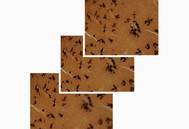
Z-stack
Full focus
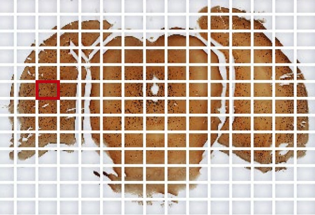
Image stitching
Objective lens: CFI Plan Apo λ 10x
Image stitching: 16 images x 13 images
Accurate counting and measurement using the same image
The clear images captured by the BZ-X not only allow for observation, but also quick quantitative measurement and analysis using the same images.
The Hybrid Cell Count function specifies an entire section as a mask area and extracts and quantifies certain cells in this area based on differences in color and luminance. With this function, the proportion of nerve cells in the entire brain section can be automatically calculated and the data can be output in spreadsheet format. This function also has a phase contrast mode that extracts and counts cultured cells without being affected by uneven luminance in the background, allowing for accurate quantitative analysis even of cultured nerve cells.
Additionally, measurement time can be significantly decreased by using Macro Cell Count to process a batch of images based on conditions extracted with Hybrid Cell Count. Conventional manual quantitative analysis requires a lot of time and effort and presents the risk that measurement conditions may vary from operator to operator. Using Hybrid Cell Count quickly provides highly reliable data with no errors caused by varying conditions.
Versatile with a single unit
The BZ-X is equipped with a high-sensitivity, high-resolution cooled CCD monochrome camera and a darkroom, allowing for advanced fluorescence, brightfield, and phase contrast observation and analysis. The BZ-X supports multi-well plates as well as Golgi-stained brain sections with a single unit, so it can perform both clear observation of various specimens and accurate measurement and evaluation with no errors. And because only a single unit is required for observation and analysis of various specimens, the BZ-X helps save space by reducing total equipment footprint.
Using the All-in-One Fluorescence Microscope BZ-X
- When a specimen is too large to be imaged with a single field of view, image stitching can be performed to capture multiple high magnification images across the sample. The images are then seamlessly joined together, creating a single fully-focused, high-resolution image of the entire specimen.
- The Z-stack function captures multiple images at different focal positions and the full focus function creates a fully focused image by combining only the areas from each image that are at their sharpest focus.
- Hybrid Cell Count can accurately extract and count just the target cells. The proportion can be calculated and output as data in spreadsheet format.
- On the basis of the conditions extracted with Hybrid Cell Count, Macro Cell Count can be used to process a batch of images quickly.
- Capable of fluorescence, brightfield, and phase contrast observation and analysis of various specimens with a single unit, the BZ-X helps save space by reducing total equipment footprint.

