Fluorescence Microscopes
Capturing high-resolution images
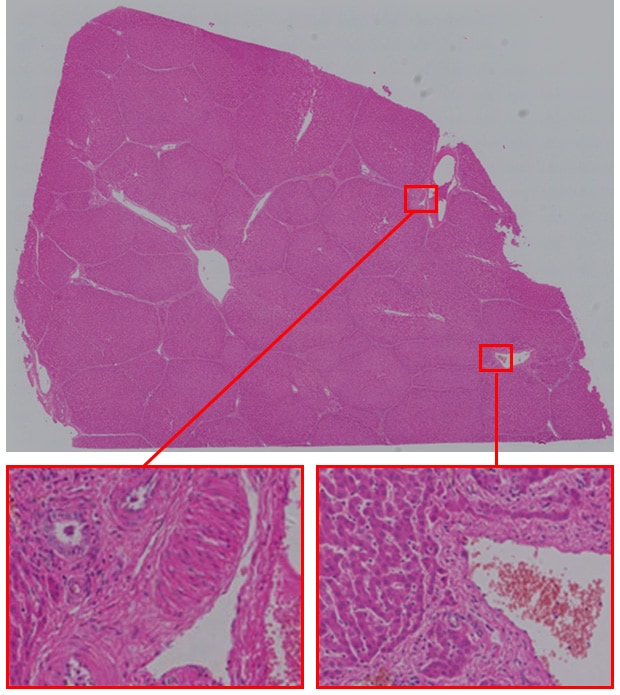
The liver has many functions. Because it plays an important role in metabolism, egestion, digestion, and other processes, medical research is underway for conditions such as fatty liver, cirrhosis, and liver cancer.
Since slices of the liver are generally large, conventional microscopes typically have to view small areas at multiple locations.
Saving at high resolution
Images can be checked and displayed at high resolution.
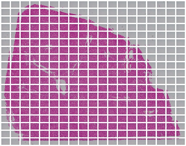
Image stitching
Objective lens: CFI Plan Apo λ 20x
Image stitching: 19 images × 19 images
Using the All-in-One Fluorescence Microscope BZ-X
- When a slice of the target is too large to fall within a single field of view, images are captured while moving the stage and a high-resolution image can be created by stitching these images.
- Even for tilted specimens or specimens that have height differences, it is possible to create an image in which the entire specimen is in focus. This is accomplished by capturing multiple images in the Z direction and stitching together only the parts that are in focus.
- An area of interest can be extracted from the whole liver and that portion can be automatically measured and analyzed with Hybrid Cell Count.
- On the basis of the conditions extracted with Hybrid Cell Count, the Macro Cell Count can be used to process a batch of multiple images.