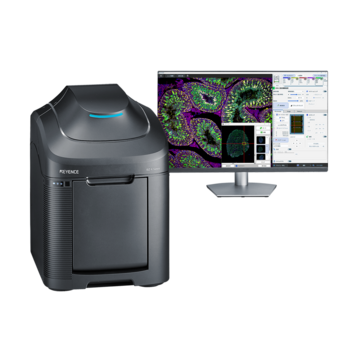
Confocal Microscope vs. Optical Microscope
Both confocal and optical microscopes are valuable imaging tools, but they serve different purposes. Optical microscopes are ideal for routine observation and general laboratory work, while confocal microscopes provide enhanced resolution, optical sectioning, and 3D imaging capabilities for more advanced applications. By evaluating factors such as sample type, imaging requirements, and budget, researchers can determine which microscope is best suited for their work.
Key Takeaways
- Optical microscopes are widely used for general magnified imaging.
- Confocal microscopes use laser scanning and a pinhole to capture clear images of thick samples.
- Confocal systems work better for thick samples, fluorescence imaging, and depth analysis.
- Optical microscopes are typically simpler, faster, and more affordable.
- The best choice depends on sample type, resolution needs, budget, and research goals.
Choosing between a confocal microscope vs optical microscope setup comes down to what you're trying to see and how deep into the sample you need to look. Both tools magnify, but they go about it in very different ways, and each fits a different kind of lab work.
What Is an Optical Microscope?
An optical microscope, sometimes called a light microscope, uses visible light and a set of lenses to magnify a sample. Light passes through or reflects off the specimen, then travels through the objective and eyepiece lenses to form an enlarged image. This basic design has been around for centuries, and it still forms the backbone of most labs, classrooms, and quality control benches.
These scopes come in many forms, from simple student models to research-grade compound microscopes, sitting alongside stereo, digital, and polarizing scopes. If you're comparing the full range of types of microscopes available, an optical model is usually the starting point, since it's affordable and easy to learn.
Optical microscopes work well on thin, flat samples. A blood smear, a tissue slice, or a printed circuit trace all show up clearly under standard light. The catch is depth. Once a sample gets thick, light scatters from areas above and below the plane you're focused on, and the image gets blurry or hazy.
What Is a Confocal Microscope?
A confocal microscope solves that blur problem with a different approach. Instead of flooding the whole sample with light, it scans a laser across one focal plane at a time. A pinhole aperture blocks out-of-focus light before it reaches the detector, so only light from the exact plane in focus makes it into the image.
The result is a sharper, cleaner picture, especially with thick or fluorescently labeled samples. Because the system builds an image plane by plane, researchers can also stack those planes into a three-dimensional reconstruction. This is one reason confocal microscopy has become a standard tool in cell biology and developmental research, where structures often extend through many layers of tissue.
How Confocal Microscopy Works
Confocal imaging is fundamentally based on point-by-point scanning as opposed to a single, broad flash of light. When a laser beam strikes a small area on the sample, the light that is released or reflected returns via the same optical route. The pinhole filters out out-of-focus light before it reaches the sensor, just where it would otherwise blur the image.
This process repeats thousands of times per image, moving the laser spot across the sample in a grid pattern. A computer then assembles all those points into a single, sharp frame. It sounds slow when described this way, but today's scanners handle it in a fraction of a second.
Types of Confocal Microscopes: Point-Scanning vs Spinning Disk Confocal Microscope
There are several types of confocal microscopes, and the two most common are point-scanning and spinning-disk systems. A point-scanning confocal microscope moves a single laser spot across the sample line by line. It tends to offer the sharpest resolution and the most flexible settings, which suits detailed, high-precision imaging.
An alternative method is used by a spinning-disk confocal microscope. It scans several places simultaneously rather than just one by using a revolving disk with several tiny pinholes. This significantly accelerates the process, which is important for imaging living cells that change or migrate over time. Although the difference has decreased as camera and disk technologies have advanced, the trade-off is often a somewhat lower resolution than that of a point-scanning system.
Choosing between the two often comes down to the sample. Fixed, static samples that require the sharpest possible detail tend to benefit from point scanning. Live, dynamic samples that require speed and low light exposure tend to favor spinning disk.
When to Use a Confocal Microscope
When the sample is dense, stacked, or tagged with fluorescent markers, a confocal arrangement makes sense. It is used in cell biology labs to monitor proteins within live cells. It is used by developmental biologists to chart the evolution of embryos.
Fluorescence work in particular benefits from confocal imaging since the pinhole cuts down on background light from out-of-focus fluorescent signals. For labs already working with fluorescent labeling, pairing a fluorescence microscope setup with confocal scanning often gives the clearest possible view of labeled structures.
Confocal systems do come with tradeoffs. They cost more, they take longer to capture an image, and they require more training to operate well. For routine checks or simple magnified views, that extra complexity may not be worth it.
When to Use an Optical Microscope
When affordability, ease of use, and speed are more important than depth, an optical microscope makes sense. Some samples don't require the level of imaging that confocal microscopes offer - those samples can be easily viewed and imaged with optical microscopes.
Cost is a real factor too. A optical microscope can cost a fraction of the price of a confocal system, making it the practical choice for labs operating on tight budgets or handling high sample volumes. If you're weighing the full range of microscope types against your budget and workflow, an optical model often meets most day-to-day needs without the added cost of laser-scanning hardware.
Frequently Asked Questions
Q Is a confocal microscope better than an optical microscope?
A
Neither is better across the board. A confocal microscope gives sharper images of thick fluorescent samples, while an optical microscope is faster and more affordable for routine samples.
Q What are the main types of confocal microscopes?
A
The two main types are point-scanning and spinning disk. Point scanning offers higher resolution, while spinning disk offers faster imaging for live samples.
Q How do you know which type of confocal microscope to use?
A
Consider the sample. Fixed samples needing fine detail suit point-scanning, while live cells that move or change quickly suit spinning disk systems.
Q What are the advantages of the BZ-X1000 laser confocal unit?
A
The BZ-X1000 has a laser confocal attachment, the BZ-XLC1, which allows researchers to combine the ease of use of a fluorescence microscope with confocal imaging.