Laser Scanning Confocal Microscopes for Life Science
Key Takeaways
- Laser scanning confocal microscopes use a focused laser and a pinhole aperture to reject out-of-focus light, producing higher-contrast images than standard widefield fluorescence.
- Optical sectioning and SIM let researchers capture clean images at specific depths within cells, tissues, and other biological samples.
- A Z-stack can create a fully focused composite image or reconstructed into a three-dimensional model.
- Confocal microscopy supports applications such as cell imaging, tissue imaging, live-cell research, microplate assays, drug discovery, and other biotechnology and pharmaceutical research.
- Modular, all-in-one systems can start as a standard fluorescence microscope and add structured-illumination optical sectioning or true point-scanning laser confocal imaging as a lab's research needs grow.
What Is a Laser Scanning Confocal Microscope?
A laser scanning confocal microscope captures sharp, high-contrast images by removing light that comes from outside the focal plane. Rather than illuminating an entire specimen at once, it focuses a single point of laser light and moves that point across the sample, line by line. A pinhole placed in front of the detector blocks scattered light before it arrives, so faint fluorescent signals aren't buried under glare from surrounding tissue.
This makes laser scanning confocal microscopy especially useful for thick or densely packed samples, since standard widefield imaging tends to blur detail sitting below the surface.
How Does a Confocal Laser Scanning Microscope Work?
A confocal laser scanning microscope relies on a few components working together. Laser light travels through the optical path and lands on one spot within the specimen. Fluorescent molecules at that spot emit light, which passes back through the same optics and through a pinhole aperture. Anything outside the exact focal plane is rejected, leaving a clean, in-focus signal for the detector.
Scanning mirrors then sweep that point across the field of view, building the image pixel by pixel. Because each point is measured individually, a confocal laser scanning microscope produces cleaner results than conventional widefield fluorescence imaging, particularly when signal strength varies across the sample.
Why Use Confocal Microscopy for Biological Samples?
Try photographing a stained tissue section under a standard widefield scope, and the result is often a mess of overlapping signal, with structures above and below the plane of interest bleeding into the shot. That's the core problem confocal detection solves. Cell clusters, spheroids, and thick tissue blocks all have real depth, and whatever sits outside the plane a researcher wants to study tends to wash out the fluorescence signal that actually matters.
A laser scanning confocal microscope gets around this by isolating one depth at a time, so a faint antibody label or a weak reporter signal doesn't get lost in the glow coming from neighboring layers. Many labs start out running a standard fluorescence microscope for day-to-day work, then add confocal capability once sample thickness or signal strength starts limiting what they can actually see.
Optical Sectioning, Z-Stacks, and 3D Imaging
Instead of physically slicing a specimen, confocal and related techniques, like optical sectioning and SIM (structured illumination microscopy), capture a series of thin, in-focus images at different heights, known as a Z-stack. Optical sectioning captures a clean image from just one thin plane of focus while rejecting blur from above and below it. While structure illumination microscopy (SIM) does this computationally by projecting a patterned light and using software to isolate the signal that tracks the pattern from the blur. Point-scanning laser confocal achieves this with a scanning laser and a pinhole resulting in the best image out of the three methods.
Researchers tracking embryo development, neuron branching, or spheroid growth depend on confocal microscopy for this depth information, since flattening a sample into one image would hide most of what matters.
Life Science Applications of Laser Scanning Confocal Microscopes
Laser scanning confocal microscopes are used in many different research settings, from university labs to pharmaceutical pipelines. Common uses include cell biology, tissue analysis, drug discovery screening, biotechnology research, and quality checks during pharmaceutical development.
Since the technique produces quantifiable data, it also fits microplate-based assays, where consistency between wells matters as much as image sharpness. Picking the right confocal microscope for life science research often starts with mapping out which of these applications matter most to a lab.
Confocal Imaging for Cells and Tissues
Fixed and live cells both benefit from confocal detection, especially when a sample carries several fluorescent markers at once. Tissue sections, which often have uneven thickness and overlapping structures, become much easier to read once out-of-focus blur is stripped away. Developmental biology and neuroscience labs regularly lean on this approach to pull meaningful signal out from surrounding noise.
Confocal Microscopy for Live-Cell Imaging and Assays
Live-cell work brings its own challenge, since cells need to stay healthy throughout imaging. A confocal laser scanning microscope can track movement, division, or drug response over time without excessive light exposure damaging the sample. That makes it practical for time-lapse studies and for microplate assays used in early-stage drug discovery, where researchers need repeatable readouts across many wells at once.
What to Consider When Choosing a Confocal Microscope
Choosing a confocal microscope for life science work comes down to a handful of practical questions. How many fluorophores does the lab typically image at once, and can the system separate those channels cleanly? Is the sample sensitive to light or heat, and does the system limit exposure during longer sessions?
Ease of use matters too, particularly in shared spaces where people with different skill levels operate the same fluorescence microscope. A dependable autofocus and straightforward software save real time, especially for labs just starting out with a laser scanning confocal microscope.
All-in-One Fluorescence Imaging, With a Path to True Laser Confocal
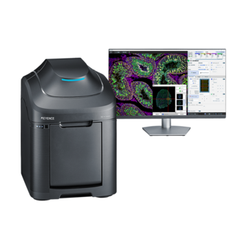
Some platforms let a lab start with a standard fluorescence microscope and add confocal capability later, rather than buying a separate dedicated system. KEYENCE's BZ-X1000 Series all-in-one fluorescence microscope includes built-in optical sectioning based on structured illumination microscopy (SIM) — a fast way to reduce out-of-focus blur in moderately thick samples using a patterned white-light source rather than a scanning laser.
Adding the BZ-XLC1 Confocal Laser Scanning Unit brings true point-scanning laser confocal to the same platform: four laser lines (405, 488, 561, and 640 nm), dual galvano scanning mirrors, an adjustable pinhole, and a high-sensitivity GaAsP detector, without requiring a separate darkroom or anti-vibration table. Labs can begin with fluorescence imaging and SIM-based sectioning, then add the BZ-XLC1 on-site once their research calls for confocal-grade resolution.
Frequently Asked Questions
Q How does a confocal laser scanning microscope work?
A
It scans a focused laser point across the sample and uses a pinhole to block out-of-focus light, building sharp images at each depth that combine into a fully focused result or a 3D dataset.
Q What is the difference between a fluorescence microscope and a confocal microscope?
A
A standard fluorescence microscope lights up the whole sample at once, while a confocal microscope isolates light from one focal plane at a time, cutting blur from regions outside that plane.
Q What is structured illumination microscopy (SIM), and how does it compare to laser scanning confocal?
A
SIM projects a patterned light source onto the sample and uses software to remove out-of-focus signal, providing optical sectioning without a scanning laser or a pinhole. It's a fast, accessible way to sharpen images of moderately thick samples; point-scanning laser confocal remains the higher-resolution option for thicker or more densely labeled specimens.
Q What are the benefits of laser scanning confocal microscopy?
A
It lowers background noise, improves contrast in thick or dense samples, and allows optical sectioning that standard widefield imaging simply can't match.
Q What biological samples can be viewed with a confocal microscope?
A
Fixed and live cells, tissue sections, organoids, and embryos are all common choices, especially when depth or fine structural detail matters.
Q Can a fluorescence microscope be upgraded to add laser scanning confocal later?
A
Yes. Modular platforms such as KEYENCE's BZ-X1000 Series can add the BZ-XLC1 Confocal Laser Scanning Unit on-site, bringing true point-scanning laser confocal imaging to an existing fluorescence microscope without a separate darkroom or vibration-isolation table.