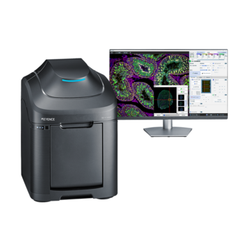
Top Confocal Microscopy Applications
Confocal microscopy has become an essential technique across life sciences and biomedical research. Its ability to capture high-resolution images, isolate specific focal planes, and generate detailed 3D reconstructions makes it valuable for a wide range of applications. As imaging requirements become more complex, confocal microscopy continues to provide researchers with deeper insights.
Key Takeaways
- Confocal microscopy is commonly used for high-resolution imaging of cells and tissues, and any sample with a lot of depth.
- Optical sectioning allows researchers to capture detailed 3D image data.
- Life science applications include cell biology, protein localization, tissue studies, and more.
- Confocal laser scanning microscopy is valuable when depth, contrast, and detail matter.
Researchers across biology, medicine, pharma, and materials science rely on confocal microscopy applications to see past the limits of standard light imaging. From tracking a single protein inside a living cell to mapping the layers of a tissue sample, this technique keeps showing up wherever depth and detail both matter.
What makes it stand out is the ability to isolate one thin plane of a sample at a time, then reassemble those planes into something researchers can actually study in three dimensions. That single feature explains why so many different fields, from cell biology to industrial inspection, have adopted it as a core imaging tool.
What is a Confocal Microscope?
A confocal microscope builds an image point by point rather than flooding the entire sample with light at once. A laser scans across the specimen, and a small pinhole blocks any light coming from outside the focal plane. Only the sharp, in-focus light reaches the detector, which is why the resulting image looks cleaner than that produced by a standard light microscope on a thick or complex sample.
This scanning approach, often called confocal laser scanning microscopy, lets researchers isolate a single thin slice of a sample at a time. Stack enough of those slices together, and you get a full 3D picture of something that would otherwise look like a blurry mess under regular light.
What is a Confocal Microscope Used For?
The list of confocal microscope uses spans a lot of ground. Biologists use it to watch proteins move inside living cells. Pathologists use it to study tissue structure in detail.
What ties these uses together is depth. Any time a sample has structure beneath the surface, whether that's a cell, a tissue block, or a machined part, a laser confocal microscope can reveal detail that a flat image would miss. That's the core reason this technique shows up so often across such different fields.
Life Science Applications
Confocal imaging is no longer a specialized technique but rather a common tool in the biological sciences. Since many biological concerns revolve around where something is located and how it moves over time, labs that investigate development, disease, or fundamental cellular function frequently base their imaging processes on these factors.
Cell Biology and Live Cell Imaging
Confocal systems are used by cell biologists to observe events in real time. Specific proteins are marked with fluorescent tags, and the confocal scan determines their precise location within the cell, whether at the membrane, in the nucleus, or somewhere in between. Compared to a typical fluorescence setup alone, the resulting images have much improved contrast because the pinhole eliminates background illumination.
Live cell imaging adds another layer of difficulty, since the sample keeps changing while it's being scanned. Researchers often need to balance image quality against scan speed and light exposure, since too much laser light can stress or damage living cells over a long imaging session.
Tissue Imaging and 3D Reconstruction
Confocal microscopes are widely used for tissue imaging because tissue sections often have uneven thickness and overlapping structures that create out-of-focus blur under standard widefield light — confocal detection strips that blur away, making individual structures like cells, vessels, and fibers much easier to distinguish. For 3D imaging, a confocal microscope captures a series of these clean, in-focus images at different depths through the tissue (a Z-stack), and that stack can then be combined into a fully focused composite or reconstructed into a three-dimensional model, letting researchers see how structures are arranged and connected throughout the tissue's depth rather than in a single flat plane.
Cancer research also leans on this capability. Tumor samples are rarely uniform, and a 3D reconstruction can reveal patterns of growth or invasion that a single flat slide would never show.
SIM vs Confocal
Structured illumination microscopy, or SIM, is another technique researchers sometimes weigh against confocal imaging. Both aim to produce sharper, more detailed images than standard light microscopy, but they get there in different ways, and each has its own strengths depending on the sample.
What Is SIM?
SIM works by projecting a patterned grid of light onto the sample and capturing several images as that pattern shifts. Combining those images using software produces a higher-resolution picture than a normal microscope could capture on its own. It's generally gentler on live samples than a laser scan, since it doesn't concentrate light into a single tight beam.
When to use SIM vs Confocal Microscopy
The decision is frequently based on the required level of detail and sample thickness. Because of its quickness and less exposure to light, SIM often performs best on thinner samples, such as a single layer of cultivated cells. With thicker, more complicated materials, confocal microscopy has an advantage since its optical sectioning can handle depth far better than SIM.
Access to equipment and financial resources is also important. While SIM setups are often more sophisticated, confocal systems are more accessible in shared imaging facilities. Many laboratories ultimately use both, selecting the instrument that best suits a particular experiment.
Sample preparation also factors into the decision. SIM generally works best with samples already optimized for standard fluorescence imaging, since it builds on that existing setup rather than requiring specialized mounting or staining. Confocal imaging, on the other hand, can handle a wider range of sample types without much extra prep, which is part of why it remains the more common choice in general-purpose imaging cores. Researchers new to advanced microscopy often start with confocal for this reason, then add SIM later once their imaging needs become more specific.
Frequently Asked Questions
Q What are the main applications of confocal microscopy?
A
Common uses include live cell imaging, tissue and 3D reconstruction, protein localization, and industrial surface or defect inspection.
Q Why is confocal microscopy useful in biology?
A
It removes out-of-focus light, giving sharper images of thick or fluorescently labeled samples, and it allows 3D reconstruction of complex structures like tissue or embryos.
Q When to use SIM vs confocal microscopy?
A
Use SIM for thinner, live samples where speed and gentler light exposure matter. Use confocal microscopy for thicker samples that need strong optical sectioning and depth resolution.