Fluorescence Microscopy vs Optical Microscopy
Fluorescence microscopy and optical microscopy each offer unique advantages depending on the imaging objective. While optical microscopy is well suited for general observation and routine analysis, fluorescence microscopy enables researchers to visualize specific cells, proteins, and biological structures with enhanced contrast. Understanding the differences between these techniques can help users select the most effective imaging method for their research or laboratory workflow.
Key Takeaways
- Optical microscopy uses visible light to view samples through magnification and has limited resolution.
- Fluorescence microscopy uses fluorescent labels to highlight specific structures, providing more detail of the sample.
- Fluorescence imaging provides stronger contrast for targeted biological features.
- Optical microscopy is often simpler, faster, and easier for routine observation.
- The right method depends on whether users need general viewing or targeted molecular detail.
Anyone setting up a new imaging workflow eventually runs into the same question: fluorescence microscopy vs optical microscopy, and which one actually fits the work at hand? Both techniques magnify a sample, but they rely on very different principles, and that difference shapes what each one can show you.
What Is Optical Microscopy?
When most people hear the word "microscope," the traditional configuration that comes to mind is optical microscopy. After passing through or reflecting off a sample, visible light passes through a number of lenses to create an enlarged picture. While many samples do receive a simple dye to enhance contrast, no additional stains or labels are needed.
For general observation, this approach is effective. Optical microscopy is a standard in clinical settings, teaching laboratories, and routine quality checks because a technician may place a slide under the lens and obtain an acceptable image in a matter of seconds. Resolution and clarity are the trade-offs. Optical microscopy does a good job at displaying general structure, but it has trouble separating a single protein or molecule from the rest of the image.
What Is Fluorescence Microscopy?
A distinct strategy is used in fluorescence microscopy. It employs fluorescent molecules that absorb light at one wavelength and emit it at another, rather than depending solely on reflected or transmitted light. The original excitation light is blocked by a filter, allowing only the fluorescent signal to reach the detector. The end result is a picture with annotated structures glowing brightly against a black backdrop.
What distinguishes fluorescence microscopy is this labeling process. By using a fluorescent marker to identify a particular protein, organelle, or even a single gene product, researchers may track the precise location of that structure within a cell or tissue sample. Even in a packed, hectic sample, the target is plainly visible since nothing else in the frame illuminates in the same manner.
Key Differences Between Fluorescence and Optical Microscopy
The core difference comes down to specificity versus simplicity. Optical microscopy shows you everything in the field of view at once, with contrast arising from natural differences in how light passes through different parts of the sample. Fluorescence microscopy narrows the view down to whatever has been labeled, ignoring everything else.
That difference carries through to nearly every part of the workflow, from how a sample is prepared to what kinds of questions the technique can actually answer.
Contrast and Image Detail
Optical microscopy relies on natural contrast, which comes from differences in density, thickness, or color across the sample. This works fine for structures with clear physical boundaries, like cell walls or tissue layers, but it falls short when trying to pick out one specific molecule among thousands of similar-looking ones.
Fluorescence imaging solves that problem directly. Because only labeled structures emit light, the background stays dark and the signal of interest pops out with sharp contrast. This makes it far easier to spot faint or small structures that would simply blend into the background under standard fluorescence vs optical microscopy comparisons based on plain visual inspection.
Sample Preparation Requirements
Optical microscopy samples are usually quick to prepare. A slide, maybe a basic stain, and you're ready to look. Fluorescence samples take more work. Researchers need to apply fluorescent antibodies, dyes, or genetically encoded markers, and each of these steps requires time and careful handling to avoid damaging the sample or producing a weak signal.
Live samples add another wrinkle. Fluorescent labeling and the light exposure required to excite those labels can stress living cells, so researchers often need to balance image quality against the amount of light and time the sample can tolerate before showing signs of damage.
Common Research Applications
Optical microscopy still covers a lot of ground in labs that don't need molecular specificity. Routine cell counts, tissue morphology checks, and general quality inspections all run fine on a standard light setup. It's fast, affordable, and doesn't require specialized reagents.
Fluorescence microscopy earns its place in more targeted work. Protein localization studies, gene expression tracking, and immunofluorescence assays all depend on the labeling step that optical microscopy simply cannot provide. Anyone working through the basics for the first time can check KEYENCE's beginner's guide to fluorescence microscopy for a walkthrough of how labeling and imaging fit together.
Advantages of Optical Microscopy
Optical microscopy holds a few clear advantages. It's faster to set up, cheaper to run, and doesn't require special reagents or dark-room imaging conditions. Samples can often be reused or re-examined without concern about fluorescent signal fading over time, as occurs with many fluorescent dyes after repeated light exposure.
It also demands less specialized training. A new lab member can learn to operate a standard light microscope in a short session, while fluorescence work usually takes more practice to get labeling, exposure, and filter settings right.
Advantages of Fluorescence Microscopy
Optical microscopy just cannot match the level of specificity provided by fluorescence microscopy. By labeling a single protein or structure, scientists can monitor its precise location, movement, and changes in distribution under various circumstances. Because of this, it is extremely useful for researching activities that occur at a molecular size, which is far smaller than what can be resolved by conventional contrast techniques.
It also supports multicolor imaging, in which multiple structures are tagged with different fluorescent markers and imaged together in a single frame. This lets researchers study relationships between structures, such as whether two proteins colocalize within a cell, in ways that optical microscopy alone cannot show. Teams running into image quality issues can also check KEYENCE's troubleshooting tips for clearer fluorescence images, which cover common causes of weak or noisy signal.
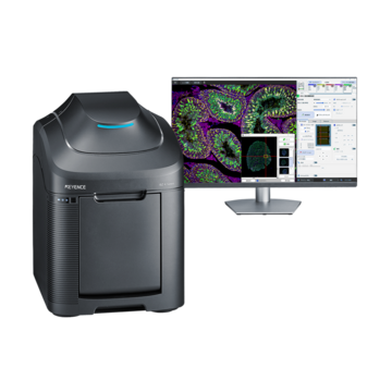
For labs weighing whether to add fluorescence capability, a dedicated fluorescence microscope setup often pays off quickly once a project moves beyond general observation and into targeted molecular work.
Frequently Asked Questions
Q What is the difference between fluorescence microscopy and optical microscopy?
A
While fluorescence microscopy employs tagged markers to highlight specific structures with high contrast against a dark background, optical microscopy uses natural light and contrast to display the overall structure of the material.
Q Is fluorescence microscopy a type of optical microscopy?
A
Yes, in a broad sense, because visible light is still used. However, it differs from conventional optical microscopy in that it uses filtered light and fluorescent tagging instead of only transmitted or reflected light.
Q When should fluorescence microscopy be used?
A
Use it when a specific protein, organelle, or molecule needs to be isolated and tracked within a sample, especially in research involving gene expression, protein localization, or live cell imaging.
Q What are the limitations of fluorescence microscopy?
A
It is slower and more expensive than ordinary optical microscopy because it involves additional sample preparation, specialized reagents, and careful control of light exposure to prevent photobleaching or damage to living material.