Digital Microscopes
Observation of Catheters Using Digital Microscopes
This section introduces examples of how observations of catheters and stents can be performed, which are common observation targets for digital microscopes.
A lot of medical equipment is used directly on human bodies, so it must provide high levels of safety and functionality.
What is a Catheter?
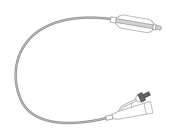
Catheters are soft tubes used for medical treatment. They are inserted into body cavities (including thoracic and abdominal cavities), lumens (including digestive and urinary tracts), or blood vessels to discharge body fluids or infuse medical solutions or contrast media.
Get detailed information on our products by downloading our catalog.
View Catalog

What is Catheter Intervention?
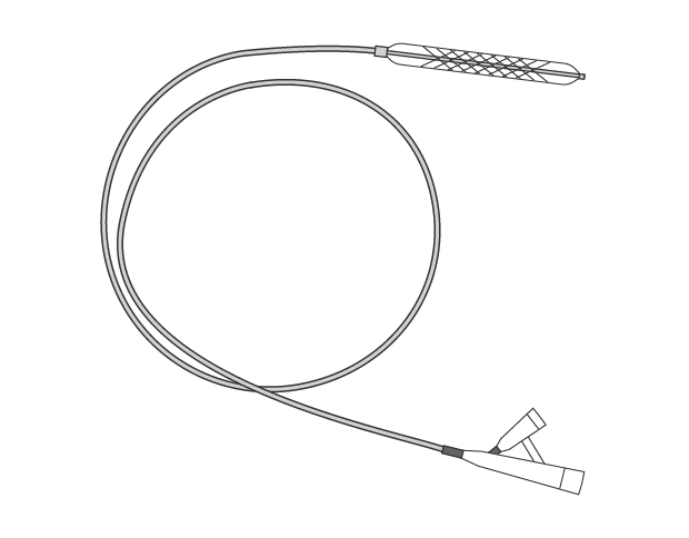
Catheter intervention expands blood vessels that are affected by arteriosclerosis and restores blood flow to its previous state. Typical catheters are usually balloon or stent types.
Balloon type catheter
A balloon type catheter is inserted along a guide wire to expand blood vessels.
The balloon applies pressure to an area affected by arteriosclerosis and expands the blood vessel to secure enough space.
(1) Run a guide wire.
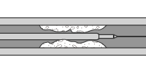
(2) Insert a balloon.
(3) Inflate the balloon.
(4) Deflate and remove the balloon.
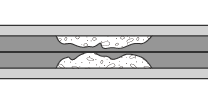
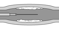
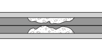
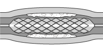
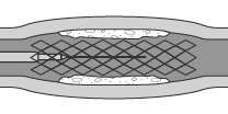
Stent type catheter
Balloon type catheters may damage the area with arteriosclerosis or the blood vessel may be blocked again. Therefore, after using a balloon type catheter, a stent type is often used as a follow up. When a stent type is used, a guide wire runs through the area with arteriosclerosis and a stent (a metal mesh tube) is placed to cover the balloon. After the balloon expands the blood vessel, it is removed, then the stent expands to support the widened blood vessel to ensure the blood vessel width.
(1) Run a guide wire.
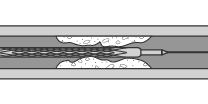
(2) Insert a balloon type catheter covered with a stent.
(3) Inflate the balloon and crimp the stent to the vessel wall.
(4) Deflate and remove the balloon.

Observation and Capturing Examples of Catheters and Stents Using Digital Microscopes
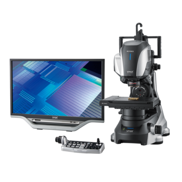
These are the latest examples of observation and capturing of catheters and stents using KEYENCE’s VHX Series 4K Digital Microscope.
Capturing and observation examples of catheters
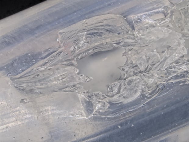
The HDR function emphasizes the surface texture of transparent objects.
Observation of flaws on a catheter surface

50×, ring illumination (HDR image)
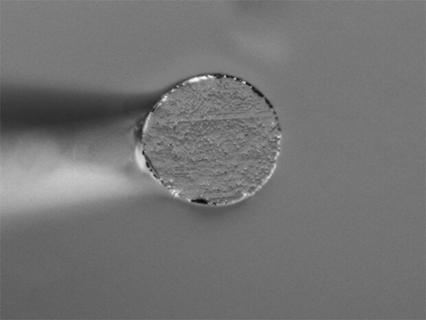
Surface observation of a catheter tip
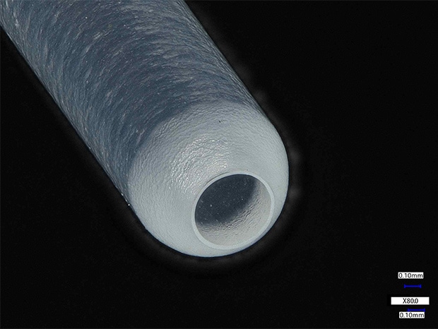
80x, ring illumination
Observation of the tip of a catheter guide wire
Optical Shadow Effect Mode allows for observation of microscopic shapes on the surface.
500×, ring illumination
500×, Optical Shadow Effect Mode image
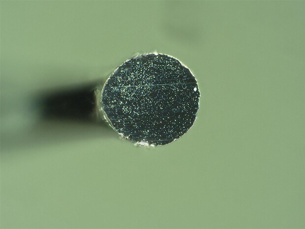
Capturing and observation examples of stents
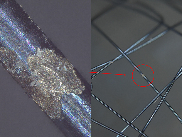
The depth composition function allows for easy focusing even at high magnification.
Stitched stent
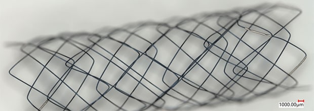
20×, mixed illumination
The image stitching function allows observation over a wide field of view with high resolution.
High magnification observation of stent surfaces
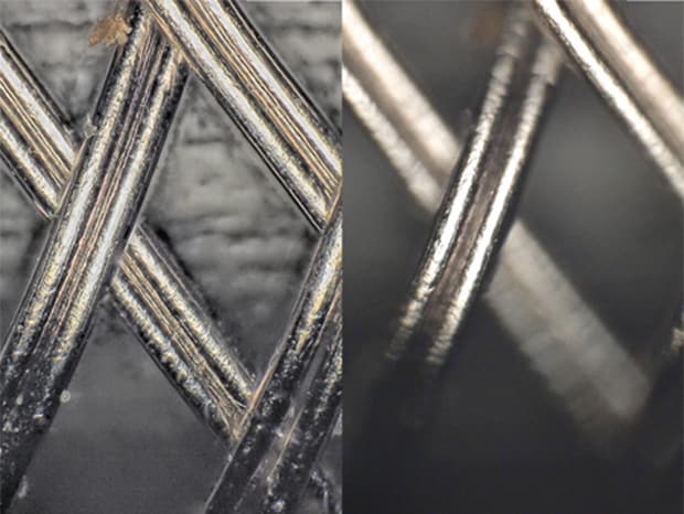
500x, coaxial illumination
High magnification observation of stent surfaces
400×, mixed illumination
Get detailed information on our products by downloading our catalog.
View Catalog