Digital Microscopes
Compound Microscope
Key Takeaways
- Compound microscopes are used to magnify small specimens using multiple lenses, making them essential for detailed observation in laboratories and research settings.
- Compound light microscopes are ideal for viewing biological samples, cells, and microorganisms with transmitted light.
- They are widely used across education, healthcare, and scientific research because of their versatility and precision.
- Choosing the right compound microscope depends on magnification needs, sample type, and intended application.

This guide explores how compound microscopes achieve high magnification through multi-lens systems and where they deliver the most value. You'll learn what distinguishes compound light microscopes from other platforms, discover applications across different fields, and understand the key factors that determine which system fits your laboratory needs.
Understanding the Basics of Compound Light Microscopes
What Is a Compound Microscope?
Compound microscopes use a series of lenses to magnify an image of an object. A specimen is collected and positioned on the stage directly underneath the objective lens, which is the one closest to the sample. A high-powered light source, sometimes located underneath the stage, illuminates the specimen. The objective lens spreads the light out so that the image appears larger. Additional lenses inside the optical path magnify the image even further. Compound light microscopes are used for a variety of applications to observe and inspect multiple types of samples, including:
- Biological: Primarily used to observe cells, tissues, and other types of biological specimens
- Metallurgical: Used to observe cross-sections of metal targets to inspect for defects and other purposes
- Polarized illumination: Features a filtering crystal that focuses light in a fixed direction to reduce reflections on the specimen's surface for a clearer image
- Phase contrast imaging: Used for colorless and transparent specimens, this instrument enhances contrast for easier viewing
The compound microscope gets its name from the compound lens system that creates magnification. Unlike simple magnifying glasses that use a single lens, these instruments stack multiple optical elements to achieve much higher magnification levels. The objective lens closest to the specimen creates an initial magnified image. The eyepiece or ocular lens then magnifies that image again, multiplying the total magnification.
What is light compound microscope technology based on? From the bottom, light passes upward through the specimen, objective lens, and then proceeds via the optical route to the camera or eyepiece. This method is ideal for thin, translucent samples such as microbe cultures, tissue sections, or blood smears. It is not necessary for the specimen to produce light. It just needs to let in enough light so that the optical system can produce detail and contrast.
What you perceive is greatly influenced by sample preparation. Too much light is blocked by thicker specimens, resulting in hazy, dark photos. For this reason, biology labs use specialized cutting instruments known as microtomes to cut tissue into slices that are only a few micrometers thick. Another level of detail is added by staining. By binding to particular cellular structures, dyes produce color contrast that draws attention to characteristics that would not be obvious in unstained samples.
These prepared specimens are transformed into precise images that show crystal formations, bacterial morphologies, or cellular architecture using a compound light microscope.
Applications Across Industries and Research
Educational institutions rely heavily on compound microscopes for teaching biology and life sciences. Students learn fundamental microscopy skills by examining prepared slides of plant cells, animal tissues, and protozoans. The hands-on experience of focusing, adjusting illumination, and identifying structures builds scientific observation skills that textbooks alone cannot provide.
Another significant application area is clinical diagnostics. Every day, compound light microscopes are used in hospital pathology labs to evaluate specimens such as tissue biopsies and blood samples. Technicians count blood cell types, identify aberrant cells, and look for bacteria or parasites.These routine examinations influence treatment decisions for millions of patients.
Applications for research are very diverse. To identify species, microbiologists look at cell and colony morphology. Botanists use cross-sections to examine the anatomy of plants. Materials scientists examine mixtures of polymers and fiber architectures. Hair, fibers, and traces of evidence are examined in forensic labs. Compound microscopy is also useful for manufacturing quality control; pharmaceutical companies use it to check tablets for contamination and coating consistency.
Differences Between Types of Microscopes
Comparing compound microscopes to stereoscopic models reveals distinct strengths. Stereo microscopes provide lower magnification, typically maxing out around 50x, but provide true three-dimensional viewing. This setup suits circuit board assembly or dissection work. Compound systems sacrifice that 3D view for much higher magnification, reaching 1000x or beyond.
Electron microscopes use electron beams to achieve magnification far beyond light-based optics. Samples require vacuum chambers and special conductive coatings. The preparation complexity and cost put electron microscopy in a different category. For samples within the resolution limits of light microscopy, compound microscopes offer a more accessible solution.
Metallurgical microscopes flip the illumination direction. Instead of light passing through specimens from below, reflected Light microscopes shine illumination down onto opaque samples. This configuration suits metal samples, ceramics, and other materials that don't transmit light.
Choosing the Right Compound Microscope
Magnification requirements drive many purchasing decisions. Teaching labs examining basic cell structure might find 400x adequate. Research applications often push to 1000x using oil immersion objectives for bacterial identification. Consider your typical specimens and what features you need to resolve.
Illumination quality affects image clarity as much as the optics themselves. Better instruments incorporate Koehler illumination systems that deliver uniform, adjustable light. LED light sources have largely replaced halogen bulbs, offering longer life and more consistent color temperature.
Financial constraints often require trade-offs between features and quality. Although entry-level compound microscopes may cost only a few hundred dollars, they often compromise on optical quality. For serious laboratory work, mid-range devices cost several thousand dollars. Align the investment with the intended use of the microscope. Simple, long-lasting systems are necessary for a teaching lab. Superior optics are necessary for a research team to publish microscope photographs.
Ergonomics is equally important. Poor posture causes neck and back strain in operators who spend hours every day on their microscope. Light microscopes with adjustable eyepiece angles and comfortable stage heights reduce fatigue.
KEYENCE’s VHX Series Digital Microscope
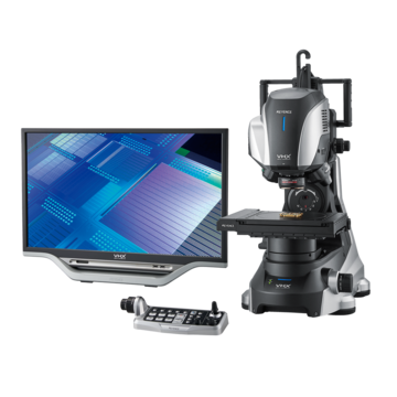
KEYENCE’s VHX Series Digital Microscope is preferred by many compound microscope users because it features some of the most advanced technology found anywhere in the world. The VHX Series digital microscope includes multiple lenses with high-power magnification capabilities ranging from 20x to 6000x. Unlike a conventional monocular compound microscope or binocular compound microscope, KEYENCE's microscopes do not feature eyepieces. Instead, the image is output directly to a digital screen for easier viewing and communication.
The versatility of the VHX is indispensable in a variety of laboratories and other environments. In addition, their ability to produce images of target samples in 4K ultra-HD resolution means they can deliver the highest-quality imaging. Plus, because they are fully automated, users don't have to waste time adjusting the focus or locating targets.
Optical Shadow Effect Mode

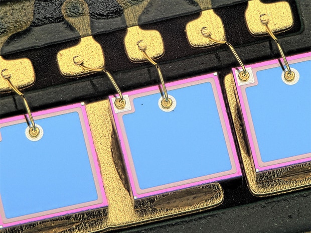
Visualize subtle surface irregularities at low magnification with Optical Shadow Effect Mode.
Fully-Automated System
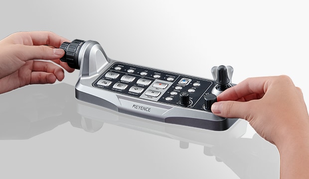
Automatic XYZ for smooth & intuitive observation at your fingertips
Fully-Integrated Head

Automatic observation from 20x to 6000x with a motorized turret

The KEYENCE Difference
When you buy a microscope from KEYENCE, you gain an instrument that goes above and beyond what you can find from other manufacturers. For example, in addition to the standard manual adjustment knobs, the VHX Series Digital Microscope comes equipped with fully automated adjustment systems that make it easier to locate and focus on your target. In addition, our high-performance illumination technology enables users to take advantage of various lighting effects and brightness levels. The included external viewing screens also make it convenient and easy to see images up close and share them with others.