3D Optical Profiling Microscope
Confocal Microscopy: Principles, Applications, and Technologies
Key Takeaways
- Confocal systems use point illumination combined with a pinhole to block out-of-focus light for high-contrast optical sectioning.
- Life science vs. industrial confocal are fundamentally different tools with the same optical principle. Life science systems optimize for fluorescence sensitivity and multi-channel detection; industrial systems optimize for calibrated z-accuracy and traceable surface metrology.
- Non-contact 3D measurement is an industrial confocal's edge, particularly on steep slopes, rough surfaces, and internal features (holes, slots) where white light interferometry struggles.
- Life science confocal systems provide sharper, higher resolution images compared to widefield microscopes and allow for imaging of thicker tissues.
- Confocal is a platform, not a single instrument. Point-scanning, spinning disk, fluorescence, and reflectance modes serve radically different use cases.
Confocal microscopy is one of the most powerful optical imaging technologies ever developed – but not all confocal systems are created for the same purpose. This guide will go into detail about what a confocal microscope is, the different types, and how to choose the right system for your work.
What Is a Confocal Microscope?
A confocal microscope is an advanced optical imaging system that uses point illumination and a spatial filter (a pinhole) to produce high-resolution, high-contrast images of a sample. The term confocal refers to the fact that the illumination point and the detector pinhole are both focused on the same point in the sample, sharing the same focal plane. This is what gives the technique its name and its power.
Traditional widefield optical microscopes illuminate an entire sample at once. Light reflects, scatters, and in fluorescence applications, fluoresces from planes above and below the focal point, reaching the detector and producing a blurry image with reduced contrast that obscures fine details. This is acceptable for thin, simple samples, but for complex, three-dimensional samples it becomes a serious limitation.
Confocal microscopes directly address this limitation by using a pinhole to block out-of-focus light before it reaches the detector. Because only one point is illuminated and detected at a time, the full image is built up by scanning the light across the sample. This enables precise optical sectioning and the ability to capture detailed structures in both 2D and 3D.
Confocal microscopy is widely used across a range of fields, from life sciences and biological research to materials science and industrial inspection, because it allows users to observe details that cannot be clearly seen with conventional optical microscopes. Whether you are imaging fluorescently tagged neurons in a living cell culture or measuring nanometer-scale surface roughness on a precision-machined part, the underlying optical principle is the same.
How Does a Confocal Microscope Work?
Confocal microscopes incorporate a pinhole aperture in front of the light-receiving element to block out-of-focus light returning from planes above and below the focal point. Only light from the precise focal plane reaches the detector, enabling sharp, high-contrast imaging. This article explains the confocal principle in four steps.
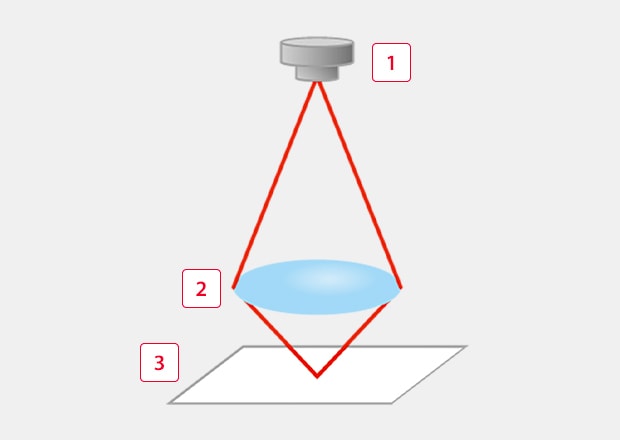
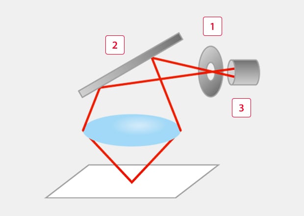
1. Light Transmission
The light source illuminates the sample through an objective lens, focusing light onto a single point at the desired focal depth. Most confocal systems use laser light sources because lasers provide coherent, monochromatic light that can be focused to a diffraction-limited spot. Laser wavelengths can be selected to match specific fluorescent dyes or to optimize surface reflectance depending on the application. Galvanometer scanning mirrors direct the focused beam across the sample point by point in the X and Y axes, systematically building up coverage of the entire field of view.
-
1Light source
-
2Objective lens
-
3Target
2. Returning Light
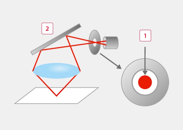
Light returning from the illuminated point travels back through the objective lens and is directed toward the detector by a beam splitter. In reflectance-based systems, used commonly in industrial and materials science applications, this is the light reflected directly from the sample surface. In fluorescence-based systems, common in life science applications, the sample emits light at a longer wavelength than the excitation source. In these cases, a dichroic mirror is used in place of a standard beam splitter — it selectively reflects the excitation wavelength toward the sample while transmitting the longer-wavelength emitted light toward the detector, separating the two signals effectively.
-
1Pinhole
-
2Half mirror
-
3Light receiving element
3. Spatial Filtering Through the Pinhole
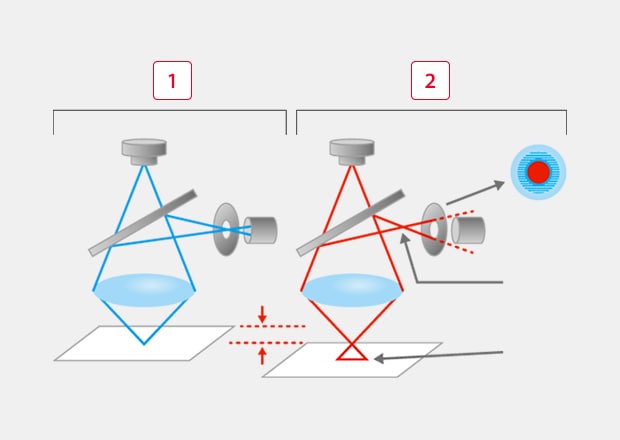
Before reaching the detector, the returning light passes through a pinhole aperture positioned at a conjugate focal plane, which means it is optically matched to the illuminated point on the sample. Light originating from the focal plane converges cleanly through the pinhole. Light originating from planes above or below the focal point converges at a different position and is largely blocked by the pinhole, preventing it from reaching the detector.
-
1Concentrated laser light
-
2Half mirror
4. Image Formation and Optical Sectioning
The light that passes through the pinhole is detected and recorded as a single data point corresponding to one scanned position on the sample. As the scanning mirrors sweep the beam across the full field of view, these data points are assembled into a complete, high-contrast 2D image of the focal plane. By stepping the focal plane incrementally along the Z-axis and acquiring a 2D image at each depth, the system captures a series of optical sections that can be reconstructed into a precise three-dimensional representation of the sample.
-
1When focused
-
2When out of focus
Types of Confocal Microscopes
While all confocal microscopes share the same fundamental principle, there are different types designed for specific applications.
Laser Confocal Microscopes for Life Science Applications
A laser confocal microscope is primarily used in biological and medical research. By using fluorescent dyes to tag cells or proteins to label structures within a sample, researchers can visualize cells, tissues, and microorganisms with exceptional clarity.
Typical applications include:
- Cell and tissue imaging
- Live-cell analysis and time-lapse imaging
- Drug development and biomedical research
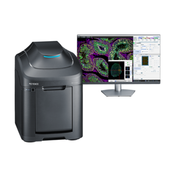
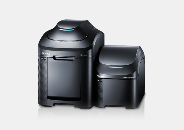
For these applications, systems like KEYENCE’s fluorescence microscopes combine high-resolution imaging with automated workflows, enabling fast and repeatable data acquisition for biological samples. The BZ-X1000 Series Fluorescence Microscope is capable of not only fluorescence, phase contrast, and brightfield imaging but with the confocal laser scanning unit, users can capture clear images without blurring for those complex and three-dimensional samples.
Laser Confocal Microscopes for Materials Research
A laser confocal microscope is commonly used in industrial and materials science applications, where precise surface measurement and profiling are required. These systems use laser-based scanning to measure surface height, roughness, and shape with high accuracy.
Often referred to as industrial confocal microscopes, they are used for:
- Surface roughness measurement
- 3D profiling of components
- Inspection of metals, semiconductors, and engineered materials
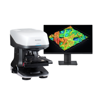
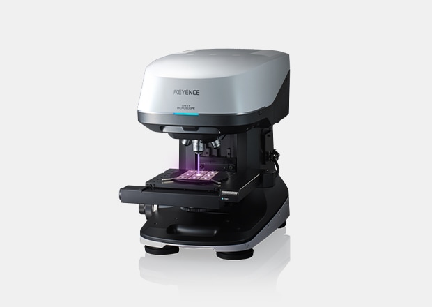
For example, systems like the KEYENCE 3D Optical Profiling Microscope integrate laser confocal technology with white light interferometry and other measurement methods to capture accurate surface data across a wide range of materials and geometries.
Need Help Figuring Out Which Microscope Is Right for You?
| I am a... | I need to... | Go to... |
|---|---|---|
| Biologist, cell scientist, neuroscientist, pathologist, or life science researcher | Image cells, tissues, organelles, or living organisms — often using fluorescent labels | Laser Confocal Microscopy for Life Science Applications |
| Engineer, materials scientist, quality control specialist, or product developer | Measure surface texture, topography, roughness, film thickness, or microstructure on physical components | Laser Scanning Confocal for Industrial & Materials Applications |
Comparing the Two Technologies
| Life science laser confocal | Industrial / Materials confocal | |
|---|---|---|
| Primary output | Fluorescence images (multi-channel) | 3D height maps, surface roughness parameters |
| Detection mode | Fluorescence emission | Reflected laser light |
| Sample preparation | Fluorescent labeling (dyes, proteins, antibodies) | None required — native surface |
| Key performance metric | Sensitivity, spectral resolution, speed | Z-accuracy, repeatability, material independence |
| Laser wavelengths | Multiple (405–640+ nm) matched to fluorophores | Optimized single wavelength for surface reflectance |
| Depth information used for | 3D cell/tissue reconstruction, co-localization | Quantitative surface topography, roughness measurement |
| Standards referenced | N/A (qualitative + quantitative biology) | ISO 25178, ISO 21920, ASME B46.1 |
| Typical sample | Cells, tissue sections, embryos, organoids | Metal parts, wafers, coatings, polymers, ceramics, optics |
| Environment | Ambient or temperature/CO2-controlled incubation | Ambient industrial or laboratory |
| Dominant user | Biologists, life scientists, clinicians | Engineers, metrologists, QC teams, R&D |
Key Advantages of Confocal Microscopy
Confocal microscopes offer several advantages over traditional optical microscopy:
- High resolution and contrast by eliminating out-of-focus light
- 3D imaging capabilities through optical sectioning
- Accurate measurement and analysis of structures or surfaces
- Non-contact measurement, ideal for delicate or sensitive samples
- Versatility across applications, from biology to industrial inspection
Applications of Confocal Microscopes
Confocal microscopes are used across many industries, with specific applications depending on the system type.
Life Sciences & Research
These applications align with fluorescence laser confocal microscopy, where image clarity and ease of use are critical for researchers.
- Imaging cells, tissues, and bacteria
- Studying biological processes at the microscopic level
- Capturing fluorescence signals for quantitative analysis
Industrial & Materials Analysis
These use cases are best supported by industrial laser confocal microscopes, where precision and repeatability are essential.
- Measuring surface roughness and texture
- Inspecting defects, wear, or coatings
- Analyzing complex geometries and microstructures
Confocal Microscopy Solutions From KEYENCE
KEYENCE offers a range of microscopy solutions designed to meet both life science and industrial confocal needs:
- Fluorescence Microscopes (BZ-X1000 Series)
Designed for biological imaging, the BZ-X Series offers high-quality fluorescence observation, automated analysis, and user-friendly operation. By using the confocal laser scanning unit, users can easily capture high-definition images without fluorescence blurring. - Laser Confocal / 3D Optical Profiling Microscopes (VK-X4000 Series)
Advanced systems capable of high-resolution surface measurement, 3D profiling, and precise analysis across various materials.
By understanding your application and measurement requirements, you can select the confocal technology that best fits your workflow.
Need help finding the right solution for you?

Frequently Asked Questions
Can a Life Science Confocal Be Used for Surface Measurements?
In principle, reflectance imaging is possible on biological confocal systems, but they lack the calibrated z-measurement systems, traceable standards, and dedicated metrology software required for quantitative surface characterization to industrial standards.
Can an Industrial Confocal Be Used to Image Fluorescent Samples?
Some industrial systems can acquire fluorescence images, but they are not optimized for the sensitivity, multi-channel detection, or live-cell environmental control that life science applications require.
What Resolution Can I Expect?
For life science systems with high-NA oil immersion objectives, lateral resolution approaches ~150–200 nm. For industrial systems, lateral resolution depends on the objective used and typically ranges from ~130 nm to ~2 µm depending on field of view requirements.
Is Confocal Better Than White Light Interferometry for Surface Measurement?
Each has strengths. White light interferometers (WLI) are extremely fast, work well on smooth surfaces, and provide unmatched vertical resolution. Industrial confocal microscopes handle steep slopes, rough surfaces, and mixed materials better, and can measure inside features (holes, slots) that WLI struggle with. Many labs use both.
What is the Difference Between Point-Scanning and Spinning Disk Confocal?
Point-scanning systems scan a single focused beam across the sample — offering flexibility and high sensitivity. Spinning disk systems use an array of pinholes to image many points simultaneously, trading some optical sectioning performance for much higher speed. Point-scanning is standard in both life science research and industrial metrology.